El objetivo de esta revisión es sintetizar la evidencia actual y las recomendaciones prácticas sobre el manejo de las urgencias y los cuidados intensivos en reptiles, ofreciendo a los clínicos un marco actualizado para su estabilización y tratamiento. La revisión aborda nueve áreas clave: anamnesis y triaje; manejo y condiciones de mantenimiento; examen físico; estabilización y reanimación cardiopulmonar (RCP); monitorización; fluidoterapia; transfusión sanguínea; analgesia y tratamiento de soporte; y diagnóstico. Se destacan las características fisiológicas que diferencian a los reptiles de los mamíferos, y cómo estas adaptaciones condicionan la fluidoterapia y la estabilización en situaciones de urgencia. Se enfatiza la rehidratación gradual y controlada dentro de la zona de temperatura óptima preferida (POTZ), así como las indicaciones específicas de las vías intravenosa, intraósea y subcutánea. Asimismo, se revisa la evidencia disponible sobre el uso de analgésicos, en particular antiinflamatorios no esteroideos (AINEs) y opioides, destacando la administración intramuscular de ketoprofeno y el predominio de receptores μ y δ. Se analizan las principales pruebas diagnósticas, como las analíticas sanguíneas, las técnicas de imagen y los cultivos en el contexto de la termorregulación. En el futuro, las investigaciones deben centrarse en ampliar los estudios farmacológicos, validar las pautas analgésicas y antibióticas y estandarizar los protocolos de monitorización y triaje para mejorar los resultados obtenidos en los cuidados intensivos en reptiles.
Abstract
The objective of this review is to summarize current evidence and practical approaches in the management of reptile emergencies and critical care, offering clinicians an evidence-based framework for stabilization and treatment. The review focuses on nine key areas: history and triage, husbandry assessment, physical examination, stabilization and cardiopulmonary resuscitation (CPR), monitoring, fluid therapy, blood transfusion, analgesia and supportive care, and diagnostic testing. The physiological characteristics that distinguish reptiles from mammals are highlighted, along with their influence on emergency stabilization and fluid therapy. Controlled, gradual rehydration within the preferred optimal temperature zone (POTZ) is emphasized, along with specific indications for intravenous, intraosseous, and subcutaneous routes. Evidence for analgesic use, particularly non-steroidal anti-inflammatory drugs (NSAIDs) and opioids, is reviewed, with intramuscular ketoprofen appearing to be a suitable option based on current evidence and with μ- and δ-receptor predominance explaining interspecies variation in opioid response. Diagnostic priorities, including blood analysis, imaging, and culture techniques, are discussed in the context of thermoregulation and stabilization. Future research should focus on expanding pharmacological studies, validating analgesic and antimicrobial regimens, and standardizing monitoring and triage protocols to improve outcomes in reptile critical care.
La presencia de este logo en un artículo de la revista indica que se publicará un examen sobre el mismo en la plataforma AVEPA Elearning. Su resolución aporta 0,15 créditos dentro del sistema de acreditaciones de especialidades veterinarias de AVEPA.
Introducción
Las urgencias en reptiles representan un reto clínico debido a sus particularidades fisiológicas, metabólicas y de manejo, que condicionan tanto la presentación clínica como la respuesta al tratamiento. El objetivo de este artículo es revisar de forma práctica el abordaje inicial, la estabilización y los cuidados intensivos en reptiles, integrando aspectos clave como el triaje, la evaluación del manejo, la fluidoterapia, la analgesia, la monitorización y las principales pruebas diagnósticas.
Anamnesis y triaje
El triaje en reptiles comienza en el momento en que el propietario contacta con la clínica. El personal de recepción y auxiliar debe estar formado para distinguir las verdaderas urgencias de aquellas situaciones que pueden esperar una cita programada sin riesgo para el paciente. Las urgencias pueden subdividirse en dos grupos: las que corresponden a procesos de inicio agudo y las que derivan de problemas crónicos con un empeoramiento repentino, ya que el abordaje, el tratamiento y el pronóstico difieren notablemente entre ambas.[Wilkinson SL: Physical examination, triage, and stabilization of the critical reptile patient. LafeberVet Webinar Library: The Critical Reptile Patient. Lafeber Company, 2023. Disponible en: https://lafeber.com/vet/the-critical-reptile-patient/, Petritz OA, Son TT: Emergency and critical care. En Divers SJ, Stahl SJ (eds): Mader’s Reptile and Amphibian Medicine and Surgery, 3ª ed., St. Louis, Elsevier, 2019; 967-976.]
La identificación precoz de casos críticos y el asesoramiento adecuado desde la primera llamada mejoran significativamente el pronóstico. Por ejemplo, la anorexia rara vez constituye una urgencia, dado que la mayoría de los reptiles toleran periodos prolongados de ayuno.[Wilkinson SL: Physical examination, triage, and stabilization of the critical reptile patient. LafeberVet Webinar Library: The Critical Reptile Patient. Lafeber Company, 2023. Disponible en: https://lafeber.com/vet/the-critical-reptile-patient/] No obstante, una anamnesis detallada ayuda a valorar la gravedad: un dragón barbudo (Pogona vitticeps) que lleva varios días sin comer, pero que se mantiene activo puede programarse para consulta no urgente, mientras que un ejemplar letárgico, débil o en decúbito lateral con los ojos cerrados requiere atención inmediata.[Wilkinson SL: Physical examination, triage, and stabilization of the critical reptile patient. LafeberVet Webinar Library: The Critical Reptile Patient. Lafeber Company, 2023. Disponible en: https://lafeber.com/vet/the-critical-reptile-patient/] De forma similar, la presencia de ruidos respiratorios leves en una tortuga puede esperar unos días; sin embargo, una respiración con la boca abierta y extensión del cuello precisa una valoración urgente.[Wilkinson SL: Physical examination, triage, and stabilization of the critical reptile patient. LafeberVet Webinar Library: The Critical Reptile Patient. Lafeber Company, 2023. Disponible en: https://lafeber.com/vet/the-critical-reptile-patient/, Petritz OA, Son TT: Emergency and critical care. En Divers SJ, Stahl SJ (eds): Mader’s Reptile and Amphibian Medicine and Surgery, 3ª ed., St. Louis, Elsevier, 2019; 967-976.]
Dado que algunos reptiles muestran comportamientos que pueden simular signos compatibles con una enfermedad, el personal debe conocer las peculiaridades específicas de cada especie para evitar errores en el triaje. Algunos ejemplos incluyen la catalepsia o inmovilidad tónica (“fingir la muerte”), en ocasiones acompañada de sangrado oral, y que puede observarse en especies como las serpientes hocico de cerdo (Heterodon spp.);[Long SY: Approach to reptile emergency medicine. Vet Clin North Am Exot Anim Pract 2016; 19(2):567-590. [PubMed], Burghardt GM, Greene HW: Predator simulation and duration of death feigning in neonate hognose snakes. Anim Behav 1988; 36(6):1842-1844., Magallón RF, Castillo M, Belton E, et al: Dead snake! A strategy for survival: Thanatosis in some Panamanian snakes with a review of death-feigning in American snakes. Reptiles Amphibians 2021; 28(3):389-396.]el movimiento de cola para simular la cabeza, y el comportamiento de señuelo caudal (caudal luring) descrito en crías de pitones arborícolas (Morelia spp.) o víboras.[Long SY: Approach to reptile emergency medicine. Vet Clin North Am Exot Anim Pract 2016; 19(2):567-590. [PubMed], Smith CF, Schuett GW: Tail movements by late-term fetal pitvipers resemble caudal luring: prenatal development of an ambush predatory behaviour. R Soc Open Sci 2022; 9(5):220218. [PubMed]] La familiaridad con este tipo de comportamientos evita alarmas innecesarias y garantiza que los casos realmente críticos reciban atención inmediata.
Durante la anamnesis se debe registrar el motivo de la consulta, la duración de los signos clínicos, la especie, la identificación del animal y cualquier atención veterinaria previa.[Wilkinson SL: Physical examination, triage, and stabilization of the critical reptile patient. LafeberVet Webinar Library: The Critical Reptile Patient. Lafeber Company, 2023. Disponible en: https://lafeber.com/vet/the-critical-reptile-patient/, Doss GA, Sladky KK: History and physical exam. En Graham JE, Beaufrère H, Doss GA (eds): Exotic Animal Emergency and Critical Care Medicine, 1ª ed., Hoboken, Wiley-Blackwell, 2021; 699-709.] También es fundamental preguntar por la conducta, la postura, el nivel de actividad y los parámetros ambientales, ya que los problemas de manejo son, con frecuencia, la causa subyacente en las consultas de urgencias.[Wilkinson SL: Physical examination, triage, and stabilization of the critical reptile patient. LafeberVet Webinar Library: The Critical Reptile Patient. Lafeber Company, 2023. Disponible en: https://lafeber.com/vet/the-critical-reptile-patient/, Doss GA, Sladky KK: History and physical exam. En Graham JE, Beaufrère H, Doss GA (eds): Exotic Animal Emergency and Critical Care Medicine, 1ª ed., Hoboken, Wiley-Blackwell, 2021; 699-709.]
Manejo y condiciones de mantenimiento
El manejo inadecuado continúa siendo la principal causa de enfermedad y presentación en urgencias de reptiles.[Wilkinson SL: Physical examination, triage, and stabilization of the critical reptile patient. LafeberVet Webinar Library: The Critical Reptile Patient. Lafeber Company, 2023. Disponible en: https://lafeber.com/vet/the-critical-reptile-patient/, Doss GA, Sladky KK: History and physical exam. En Graham JE, Beaufrère H, Doss GA (eds): Exotic Animal Emergency and Critical Care Medicine, 1ª ed., Hoboken, Wiley-Blackwell, 2021; 699-709.] Evaluar las condiciones ambientales, nutricionales y de iluminación resulta esencial tanto para la valoración clínica como para la estabilización del paciente.[Wilkinson SL: Physical examination, triage, and stabilization of the critical reptile patient. LafeberVet Webinar Library: The Critical Reptile Patient. Lafeber Company, 2023. Disponible en: https://lafeber.com/vet/the-critical-reptile-patient/, Petritz OA, Son TT: Emergency and critical care. En Divers SJ, Stahl SJ (eds): Mader’s Reptile and Amphibian Medicine and Surgery, 3ª ed., St. Louis, Elsevier, 2019; 967-976., Long SY: Approach to reptile emergency medicine. Vet Clin North Am Exot Anim Pract 2016; 19(2):567-590. [PubMed], Doss GA, Sladky KK: History and physical exam. En Graham JE, Beaufrère H, Doss GA (eds): Exotic Animal Emergency and Critical Care Medicine, 1ª ed., Hoboken, Wiley-Blackwell, 2021; 699-709.] Incluso cuando no es posible una atención inmediata, la recopilación de esta información permite al personal ofrecer recomendaciones provisionales, como ajustar la temperatura, la humedad o la exposición a radiación ultravioleta B (UVB), con el fin de mejorar la condición del animal antes de su llegada a la clínica.[Wilkinson SL: Physical examination, triage, and stabilization of the critical reptile patient. LafeberVet Webinar Library: The Critical Reptile Patient. Lafeber Company, 2023. Disponible en: https://lafeber.com/vet/the-critical-reptile-patient/, Doss GA, Sladky KK: History and physical exam. En Graham JE, Beaufrère H, Doss GA (eds): Exotic Animal Emergency and Critical Care Medicine, 1ª ed., Hoboken, Wiley-Blackwell, 2021; 699-709.]
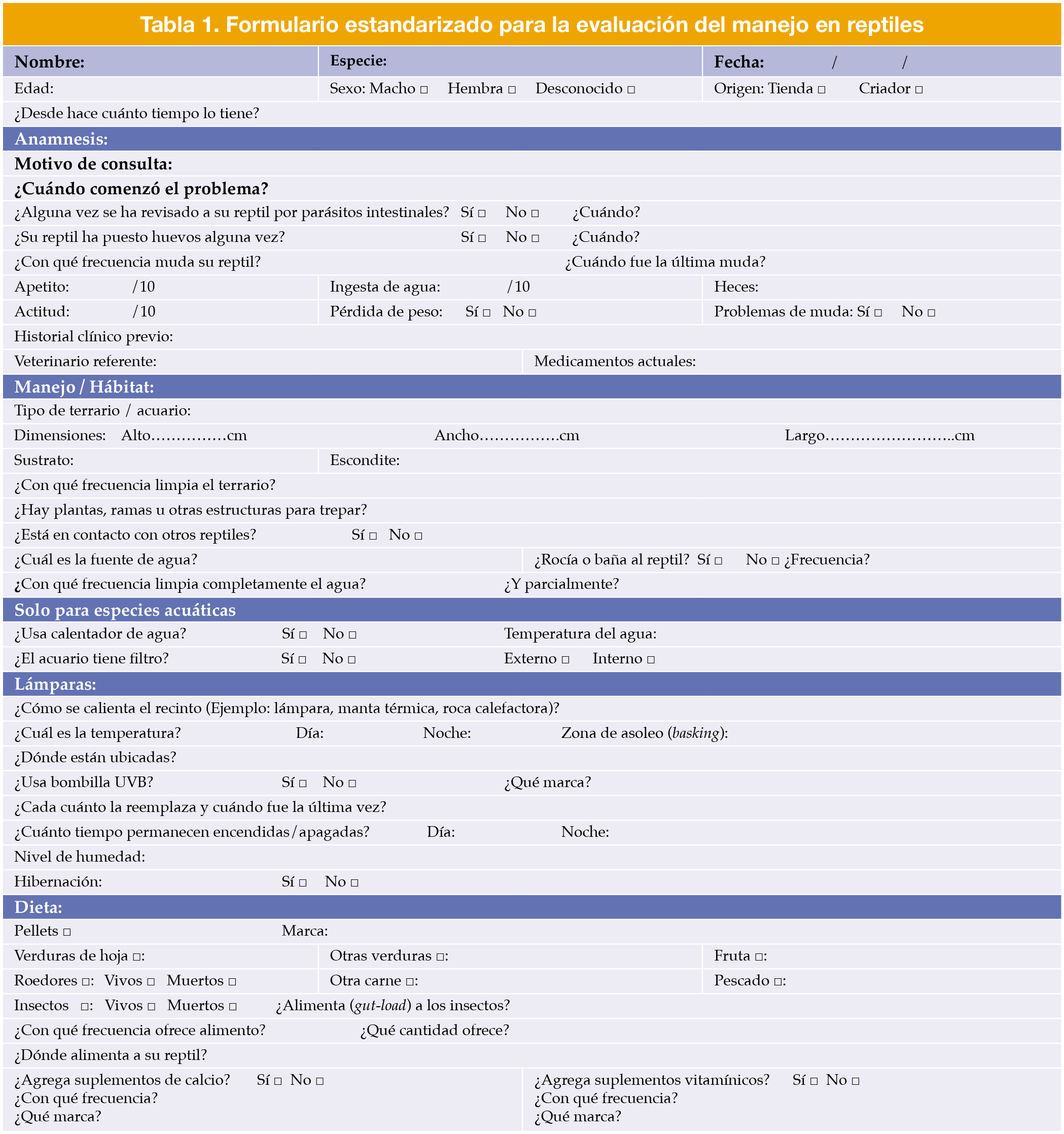
El uso de un cuestionario estandarizado facilita una evaluación sistemática del terrario, en el que se incluyen las características del recinto, los parámetros ambientales, la dieta y la calidad del agua (Tabla 1).[Long SY: Approach to reptile emergency medicine. Vet Clin North Am Exot Anim Pract 2016; 19(2):567-590. [PubMed]] Estos datos permiten distinguir las patologías derivadas de un manejo incorrecto de otras urgencias médicas.[Petritz OA, Son TT: Emergency and critical care. En Divers SJ, Stahl SJ (eds): Mader’s Reptile and Amphibian Medicine and Surgery, 3ª ed., St. Louis, Elsevier, 2019; 967-976.] En muchos casos, la corrección de los factores ambientales por sí sola puede evitar la progresión o el empeoramiento de los signos clínicos.[Wilkinson SL: Physical examination, triage, and stabilization of the critical reptile patient. LafeberVet Webinar Library: The Critical Reptile Patient. Lafeber Company, 2023. Disponible en: https://lafeber.com/vet/the-critical-reptile-patient/]
Tabla 1
Examen físico
El examen físico en situaciones de urgencia debe ser breve, sistemático y lo menos estresante posible.[Wilkinson SL: Physical examination, triage, and stabilization of the critical reptile patient. LafeberVet Webinar Library: The Critical Reptile Patient. Lafeber Company, 2023. Disponible en: https://lafeber.com/vet/the-critical-reptile-patient/] Dado que la manipulación excesiva puede provocar descompensación o alteración de los hallazgos clínicos, es fundamental preparar todo el material necesario con antelación y realizar una evaluación inicial a distancia antes de sujetar al animal.[Doss GA, Sladky KK: History and physical exam. En Graham JE, Beaufrère H, Doss GA (eds): Exotic Animal Emergency and Critical Care Medicine, 1ª ed., Hoboken, Wiley-Blackwell, 2021; 699-709.] Esta evaluación inicial se centra en el estado mental, la postura, la condición y el movimiento corporal.[Wilkinson SL: Physical examination, triage, and stabilization of the critical reptile patient. LafeberVet Webinar Library: The Critical Reptile Patient. Lafeber Company, 2023. Disponible en: https://lafeber.com/vet/the-critical-reptile-patient/, Doss GA, Sladky KK: History and physical exam. En Graham JE, Beaufrère H, Doss GA (eds): Exotic Animal Emergency and Critical Care Medicine, 1ª ed., Hoboken, Wiley-Blackwell, 2021; 699-709.] Los lagartos y quelonios suelen mantener el cuerpo elevado al desplazarse; la imposibilidad de hacerlo sugiere debilidad, disfunción neurológica o enfermedad sistémica.[Music MK, Strunk A: Reptile critical care and common emergencies. Vet Clin North Am Exot Anim Pract 2016; 19(2):591-612. [PubMed]] Debe observarse la frecuencia y el esfuerzo respiratorio antes de la manipulación, ya que esta modifica el patrón respiratorio.[Wilkinson SL: Physical examination, triage, and stabilization of the critical reptile patient. LafeberVet Webinar Library: The Critical Reptile Patient. Lafeber Company, 2023. Disponible en: https://lafeber.com/vet/the-critical-reptile-patient/, Music MK, Strunk A: Reptile critical care and common emergencies. Vet Clin North Am Exot Anim Pract 2016; 19(2):591-612. [PubMed]] La inspección visual también puede revelar signos de deshidratación (como ojos hundidos), mudas incompletas, presencia de ectoparásitos o lesiones por quemaduras o traumatismos.[Doss GA, Sladky KK: History and physical exam. En Graham JE, Beaufrère H, Doss GA (eds): Exotic Animal Emergency and Critical Care Medicine, 1ª ed., Hoboken, Wiley-Blackwell, 2021; 699-709.]
Tras la evaluación inicial sin manipulación, se realiza un examen físico dirigido que incluya la valoración del estado de hidratación, la frecuencia cardíaca (mediante Doppler o estetoscopio), la palpación celómica y una revisión neurológica y musculoesquelética básica.[Doss GA, Sladky KK: History and physical exam. En Graham JE, Beaufrère H, Doss GA (eds): Exotic Animal Emergency and Critical Care Medicine, 1ª ed., Hoboken, Wiley-Blackwell, 2021; 699-709., Music MK, Strunk A: Reptile critical care and common emergencies. Vet Clin North Am Exot Anim Pract 2016; 19(2):591-612. [PubMed]] En las serpientes se debe valorar la fuerza con la que se enrollan y la coordinación, mientras que en tortugas y lagartos, hay que evaluar el reflejo de enderezamiento (righting reflex).[Doss GA, Sladky KK: History and physical exam. En Graham JE, Beaufrère H, Doss GA (eds): Exotic Animal Emergency and Critical Care Medicine, 1ª ed., Hoboken, Wiley-Blackwell, 2021; 699-709., Music MK, Strunk A: Reptile critical care and common emergencies. Vet Clin North Am Exot Anim Pract 2016; 19(2):591-612. [PubMed]] La exploración oral debe valorar el color de las mucosas, el tiempo de relleno capilar y la viscosidad de la saliva; las mucosas pálidas pueden indicar anemia, mientras que la hiperemia o la presencia de placas pueden sugerir estomatitis.[Doss GA, Sladky KK: History and physical exam. En Graham JE, Beaufrère H, Doss GA (eds): Exotic Animal Emergency and Critical Care Medicine, 1ª ed., Hoboken, Wiley-Blackwell, 2021; 699-709.] Asimismo, en reptiles con mucosas no pigmentadas, una coloración amarillenta de las mucosas puede indicar hiperbilirrubinemia asociada a enfermedad hepatobiliar en aquellas especies capaces de producir bilirrubina (p. ej., Pogona vitticeps). Sin embargo, muchos reptiles carecen de la enzima biliverdina reductasa y producen principalmente biliverdina en lugar de bilirrubina, por lo que la ictericia es poco evidente y puede observarse con mayor frecuencia una coloración verdosa de la orina o de los uratos.[Divers SJ: Hepatology. En Divers SJ, Stahl SJ (eds): Mader’s Reptile and Amphibian Medicine and Surgery, 3ª ed., St. Louis, Elsevier, 2019; 649-668.]
El objetivo del examen físico en una consulta de urgencia es determinar si el paciente está lo suficientemente estable para continuar con las pruebas diagnósticas y una estabilización gradual, o si es una emergencia y requiere reanimación cardiopulmonar y estabilización inmediata antes de proceder con estudios adicionales.[Doss GA, Sladky KK: History and physical exam. En Graham JE, Beaufrère H, Doss GA (eds): Exotic Animal Emergency and Critical Care Medicine, 1ª ed., Hoboken, Wiley-Blackwell, 2021; 699-709.]
Reanimación cardiopulmonar (RCP) y estabilización
Los pacientes que mantienen un estado mental adecuado, el reflejo de enderezamiento y la respiración espontánea se consideran estables, mientras que aquellos con apnea, paro cardíaco o bradicardia severa requieren RCP inmediata.[Wilkinson SL: Physical examination, triage, and stabilization of the critical reptile patient. LafeberVet Webinar Library: The Critical Reptile Patient. Lafeber Company, 2023. Disponible en: https://lafeber.com/vet/the-critical-reptile-patient/, Martinez-Jimenez D, Hernandez-Divers SJ: Emergency care of reptiles. Vet Clin North Am Exot Anim Pract 2007; 10(2):557-585. [PubMed]]
Pacientes inestables – RCP
Los reptiles presentan una tolerancia excepcional a la hipoxia, gracias a la rápida transición al metabolismo anaerobio, la conversión hepática del lactato en bicarbonato y el almacenamiento de lactato cálcico en el caparazón de las tortugas.[Long SY: Approach to reptile emergency medicine. Vet Clin North Am Exot Anim Pract 2016; 19(2):567-590. [PubMed], Martinez-Jimenez D, Hernandez-Divers SJ: Emergency care of reptiles. Vet Clin North Am Exot Anim Pract 2007; 10(2):557-585. [PubMed], Kanda I, Brandão J: Oxygen therapy. En Graham JE, Beaufrère H, Doss GA (eds): Exotic Animal Emergency and Critical Care Medicine, 1ª ed., Hoboken, Wiley-Blackwell, 2021; 716-721.] Además, su ventrículo parcialmente dividido permite la existencia de shunts derecha–izquierda durante la apnea o la alteración pulmonar, manteniendo la oxigenación sistémica.[Long SY: Approach to reptile emergency medicine. Vet Clin North Am Exot Anim Pract 2016; 19(2):567-590. [PubMed]] Algunas tortugas son capaces de incrementar la expresión de proteínas de choque térmico y antioxidantes en condiciones anóxicas, lo que explica su notable capacidad de recuperación al daño cerebral o isquémico en comparación con mamíferos y aves.[Lutz P, Milton S: Negotiating brain anoxia survival in the turtle. J Exp Biol 2004; 207:314-317. [PubMed], Bickler P: Effects of temperature and anoxia on regional cerebral blood flow in turtles. Am J Physiol 1992; 262:538-541. [PubMed], Prentice H, Milton S, Scheurle D, et al: The upregulation of cognate and inducible heat shock proteins in the anoxic turtle brain. J Cereb Blood Flow Metab 2004; 24:826-828. [PubMed], Wasser J, Meinertz E, Chang S: Metabolic and cardiodynamic responses of isolated turtle hearts to ischemia and reperfusion. Am J Physiol 1992; 262:437-443. [PubMed]] Aun así, esta tolerancia no implica que la ventilación sea un factor secundario durante la reanimación, ya que la ventilación no solo permite la oxigenación, sino que también regula el equilibrio ácido-base. Dado que sus tejidos pueden tolerar una hipoxia prolongada, pero no está demostrado que toleren una acidosis metabólica sostenida, la intervención ventilatoria temprana resulta más eficaz que la compresión cardíaca inmediata.[Petritz OA, Son TT: Emergency and critical care. En Divers SJ, Stahl SJ (eds): Mader’s Reptile and Amphibian Medicine and Surgery, 3ª ed., St. Louis, Elsevier, 2019; 967-976., Martinez-Jimenez D, Hernandez-Divers SJ: Emergency care of reptiles. Vet Clin North Am Exot Anim Pract 2007; 10(2):557-585. [PubMed], Doss GA, Sladky KK: CPR and euthanasia. En Graham JE, Beaufrère H, Doss GA (eds): Exotic Animal Emergency and Critical Care Medicine, 1ª ed., Hoboken, Wiley-Blackwell, 2021; 738-745.] Debido a estas adaptaciones fisiológicas únicas, la reanimación en reptiles debe seguir la secuencia de vía aérea, respiración y circulación (ABC: Airway, Breathingy Circulation):[Petritz OA, Son TT: Emergency and critical care. En Divers SJ, Stahl SJ (eds): Mader’s Reptile and Amphibian Medicine and Surgery, 3ª ed., St. Louis, Elsevier, 2019; 967-976., Martinez-Jimenez D, Hernandez-Divers SJ: Emergency care of reptiles. Vet Clin North Am Exot Anim Pract 2007; 10(2):557-585. [PubMed], Doss GA, Sladky KK: CPR and euthanasia. En Graham JE, Beaufrère H, Doss GA (eds): Exotic Animal Emergency and Critical Care Medicine, 1ª ed., Hoboken, Wiley-Blackwell, 2021; 738-745.]
- Vía aérea y respiración: se debe intubar y comprobar la permeabilidad de la vía aérea. Iniciar ventilación con presión positiva intermitente (4-6 respiraciones/min, presión máxima <10 cm H₂O), evitando el uso prolongado del oxígeno al 100 % por su posible efecto depresor sobre la respiración.[Petritz OA, Son TT: Emergency and critical care. En Divers SJ, Stahl SJ (eds): Mader’s Reptile and Amphibian Medicine and Surgery, 3ª ed., St. Louis, Elsevier, 2019; 967-976., Martinez-Jimenez D, Hernandez-Divers SJ: Emergency care of reptiles. Vet Clin North Am Exot Anim Pract 2007; 10(2):557-585. [PubMed], Doss GA, Sladky KK: CPR and euthanasia. En Graham JE, Beaufrère H, Doss GA (eds): Exotic Animal Emergency and Critical Care Medicine, 1ª ed., Hoboken, Wiley-Blackwell, 2021; 738-745.] En reptiles, la hipercapnia aumenta el volumen corriente, mientras que la hipoxia estimula la frecuencia respiratoria, lo cual facilita la recuperación de la respiración espontánea.[Petritz OA, Son TT: Emergency and critical care. En Divers SJ, Stahl SJ (eds): Mader’s Reptile and Amphibian Medicine and Surgery, 3ª ed., St. Louis, Elsevier, 2019; 967-976., Long SY: Approach to reptile emergency medicine. Vet Clin North Am Exot Anim Pract 2016; 19(2):567-590. [PubMed], Kanda I, Brandão J: Oxygen therapy. En Graham JE, Beaufrère H, Doss GA (eds): Exotic Animal Emergency and Critical Care Medicine, 1ª ed., Hoboken, Wiley-Blackwell, 2021; 716-721.] Por este motivo, breves periodos controlados de hipoxia o hipercapnia en los que se desconecta al animal de la ventilación, pueden potenciar el estímulo respiratorio.[Martinez-Jimenez D, Hernandez-Divers SJ: Emergency care of reptiles. Vet Clin North Am Exot Anim Pract 2007; 10(2):557-585. [PubMed]]
- Circulación: las compresiones externas son anatómicamente limitadas en muchos reptiles.[Petritz OA, Son TT: Emergency and critical care. En Divers SJ, Stahl SJ (eds): Mader’s Reptile and Amphibian Medicine and Surgery, 3ª ed., St. Louis, Elsevier, 2019; 967-976.] En las tortugas, la rigidez del caparazón impide las compresiones externas.[Martinez-Jimenez D, Hernandez-Divers SJ: Emergency care of reptiles. Vet Clin North Am Exot Anim Pract 2007; 10(2):557-585. [PubMed], Doss GA, Sladky KK: CPR and euthanasia. En Graham JE, Beaufrère H, Doss GA (eds): Exotic Animal Emergency and Critical Care Medicine, 1ª ed., Hoboken, Wiley-Blackwell, 2021; 738-745.] En la mayoría de los lagartos, el corazón se localiza dentro de la cintura escapular, lo que limita la eficacia de las compresiones torácicas externas; sin embargo, los varánidos y los tegus constituyen excepciones, ya que su corazón se encuentra fuera de la cintura escapular, permitiendo una mayor eficacia de las compresiones.[Petritz OA, Son TT: Emergency and critical care. En Divers SJ, Stahl SJ (eds): Mader’s Reptile and Amphibian Medicine and Surgery, 3ª ed., St. Louis, Elsevier, 2019; 967-976., Martinez-Jimenez D, Hernandez-Divers SJ: Emergency care of reptiles. Vet Clin North Am Exot Anim Pract 2007; 10(2):557-585. [PubMed], Doss GA, Sladky KK: CPR and euthanasia. En Graham JE, Beaufrère H, Doss GA (eds): Exotic Animal Emergency and Critical Care Medicine, 1ª ed., Hoboken, Wiley-Blackwell, 2021; 738-745.] En las serpientes, deben aplicarse sobre el área cardíaca, aproximadamente en el primer tercio de la longitud corporal.[Martinez-Jimenez D, Hernandez-Divers SJ: Emergency care of reptiles. Vet Clin North Am Exot Anim Pract 2007; 10(2):557-585. [PubMed], Doss GA, Sladky KK: CPR and euthanasia. En Graham JE, Beaufrère H, Doss GA (eds): Exotic Animal Emergency and Critical Care Medicine, 1ª ed., Hoboken, Wiley-Blackwell, 2021; 738-745.]
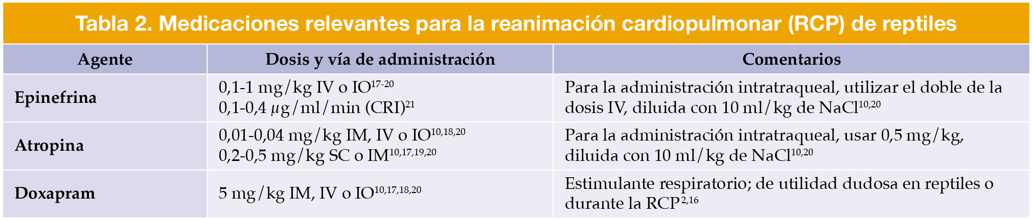
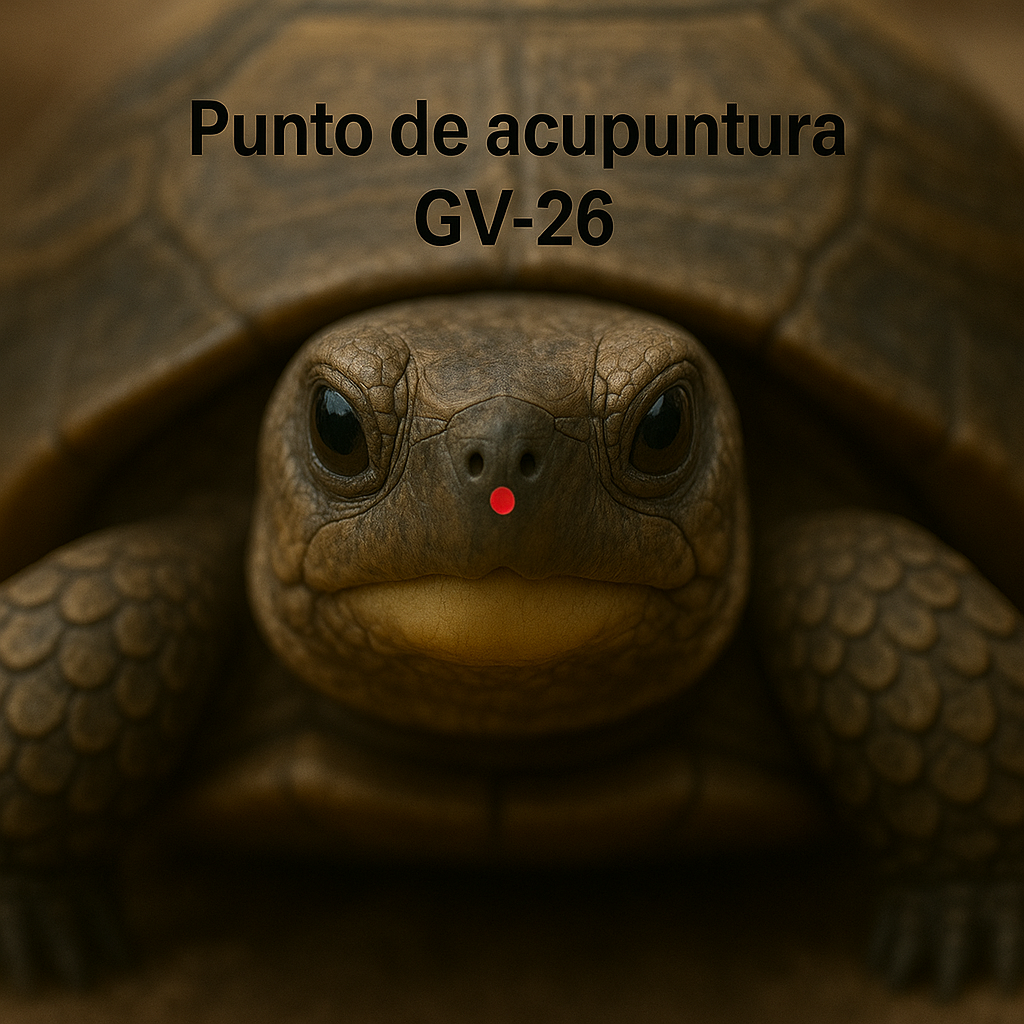
Durante la RCP, la temperatura corporal del paciente debe incrementarse gradualmente hasta su zona de temperatura óptima preferida (POTZ) y se deben administrar fluidos atemperados para mejorar la perfusión y la recuperación metabólica.[Petritz OA, Son TT: Emergency and critical care. En Divers SJ, Stahl SJ (eds): Mader’s Reptile and Amphibian Medicine and Surgery, 3ª ed., St. Louis, Elsevier, 2019; 967-976., Long SY: Approach to reptile emergency medicine. Vet Clin North Am Exot Anim Pract 2016; 19(2):567-590. [PubMed]] El soporte farmacológico debe complementar las maniobras de RCP con el fin de optimizar el resultado de la reanimación (Tabla 2).[Doss GA, Sladky KK: CPR and euthanasia. En Graham JE, Beaufrère H, Doss GA (eds): Exotic Animal Emergency and Critical Care Medicine, 1ª ed., Hoboken, Wiley-Blackwell, 2021; 738-745., Costello MF: Principles of cardiopulmonary cerebral resuscitation in special species. Semin Avian Exot Pet Med 2004; 13(3):132-141., Norton TM: Chelonian emergency and critical care. Semin Avian Exot Pet Med 2005; 14(2):106-130., Mitchell MA: Managing the reptile patient in the veterinary hospital: establishing a standard of care model for nontraditional species. J Exot Pet Med 2010; 19(1):56-72., Sladky KK, Klaphake E, Di Girolamo N, Carpenter JW: Reptiles. En Carpenter JW, Marion CJ (eds): Carpenter’s Exotic Animal Formulary, 6ª ed., St. Louis, Elsevier, 2023; 101-173., Sladky KK, Mans C: Analgesia. En Divers SJ, Stahl SJ (eds): Mader’s Reptile and Amphibian Medicine and Surgery, 3ª ed., St. Louis, Elsevier, 2019; 465-474.] Además, la estimulación del punto de acupuntura GV-26 puede inducir o favorecer la respiración espontánea y puede utilizarse como una medida de soporte complementaria durante la reanimación (Fig. 1).[Sladky KK, Mans C: Analgesia. En Divers SJ, Stahl SJ (eds): Mader’s Reptile and Amphibian Medicine and Surgery, 3ª ed., St. Louis, Elsevier, 2019; 465-474.]
Localización del punto de acupuntura GV-26 en una tortuga de tierra. Este punto se encuentra en la línea media, justo entre las narinas y el borde rostral del pico.
Pacientes inestables – Monitorización
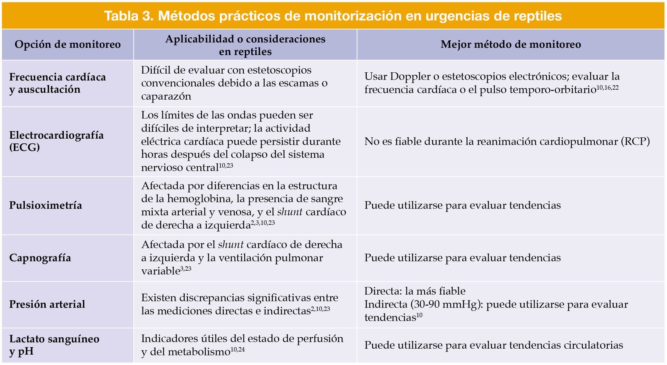
La monitorización en reptiles representa un desafío inherente debido a las notables diferencias anatómicas y fisiológicas con respecto a los mamíferos.[Long SY: Approach to reptile emergency medicine. Vet Clin North Am Exot Anim Pract 2016; 19(2):567-590. [PubMed]] Los dispositivos Doppler o los estetoscopios electrónicos continúan siendo las herramientas más prácticas para evaluar la frecuencia cardíaca y la perfusión, pudiendo colocarse sobre la región cardíaca o temporo-orbitaria para mantener libre el acceso durante las compresiones torácicas.[Martinez-Jimenez D, Hernandez-Divers SJ: Emergency care of reptiles. Vet Clin North Am Exot Anim Pract 2007; 10(2):557-585. [PubMed], Doss GA, Sladky KK: CPR and euthanasia. En Graham JE, Beaufrère H, Doss GA (eds): Exotic Animal Emergency and Critical Care Medicine, 1ª ed., Hoboken, Wiley-Blackwell, 2021; 738-745., Ito M, Levitskiy AA, Di Girolamo N: Temporoorbital pulse rate can be obtained in ball pythons (Python regius) of any size and age and agrees with heart rate. Vet Rec 2024; 195(8):e4596. [PubMed]] Los métodos convencionales, como el electrocardiograma (ECG), la pulsioximetría, la capnografía y la medición indirecta de la presión arterial suelen proporcionar datos poco fiables o inconsistentes (Tabla 3).[Petritz OA, Son TT: Emergency and critical care. En Divers SJ, Stahl SJ (eds): Mader’s Reptile and Amphibian Medicine and Surgery, 3ª ed., St. Louis, Elsevier, 2019; 967-976., Martinez-Jimenez D, Hernandez-Divers SJ: Emergency care of reptiles. Vet Clin North Am Exot Anim Pract 2007; 10(2):557-585. [PubMed], Doss GA, Sladky KK: CPR and euthanasia. En Graham JE, Beaufrère H, Doss GA (eds): Exotic Animal Emergency and Critical Care Medicine, 1ª ed., Hoboken, Wiley-Blackwell, 2021; 738-745., Ito M, Levitskiy AA, Di Girolamo N: Temporoorbital pulse rate can be obtained in ball pythons (Python regius) of any size and age and agrees with heart rate. Vet Rec 2024; 195(8):e4596. [PubMed], Doss GA, Mans C, Sladky KK: Analgesia, anesthesia, and monitoring. En Graham JE, Beaufrère H, Doss GA (eds): Exotic Animal Emergency and Critical Care Medicine, 1ª ed., Hoboken, Wiley-Blackwell, 2021; 746-757., DiGeronimo PM, Di Girolamo N: STAT diagnostics. En Graham JE, Beaufrère H, Doss GA (eds): Exotic Animal Emergency and Critical Care Medicine, 1ª ed., Hoboken, Wiley-Blackwell, 2021; 771-782.] Ante estas limitaciones, la monitorización debe centrarse en las tendencias fisiológicas más que en los valores absolutos, asegurando que las decisiones clínicas se basen en la actitud, el estado mental y los reflejos del animal.[Doss GA, Mans C, Sladky KK: Analgesia, anesthesia, and monitoring. En Graham JE, Beaufrère H, Doss GA (eds): Exotic Animal Emergency and Critical Care Medicine, 1ª ed., Hoboken, Wiley-Blackwell, 2021; 746-757.]
Tabla 3
Pacientes estables – Estabilización ambiental y fisiológica
En los reptiles estables pero comprometidos, la corrección de los parámetros ambientales constituye la primera prioridad.[Wilkinson SL: Physical examination, triage, and stabilization of the critical reptile patient. LafeberVet Webinar Library: The Critical Reptile Patient. Lafeber Company, 2023. Disponible en: https://lafeber.com/vet/the-critical-reptile-patient/] El metabolismo, la función cardiovascular y la eficacia farmacológica son óptimos únicamente dentro de la POTZ, generalmente entre 26 y 29 °C, dependiendo de la especie.[Petritz OA, Son TT: Emergency and critical care. En Divers SJ, Stahl SJ (eds): Mader’s Reptile and Amphibian Medicine and Surgery, 3ª ed., St. Louis, Elsevier, 2019; 967-976., Long SY: Approach to reptile emergency medicine. Vet Clin North Am Exot Anim Pract 2016; 19(2):567-590. [PubMed], Martinez-Jimenez D, Hernandez-Divers SJ: Emergency care of reptiles. Vet Clin North Am Exot Anim Pract 2007; 10(2):557-585. [PubMed]] Se recomienda un incremento gradual de temperatura corporal a lo largo de 4-6 horas para evitar el sobrecalentamiento.[Petritz OA, Son TT: Emergency and critical care. En Divers SJ, Stahl SJ (eds): Mader’s Reptile and Amphibian Medicine and Surgery, 3ª ed., St. Louis, Elsevier, 2019; 967-976., Martinez-Jimenez D, Hernandez-Divers SJ: Emergency care of reptiles. Vet Clin North Am Exot Anim Pract 2007; 10(2):557-585. [PubMed]] Una vez alcanzados 25-26 °C, el animal debe trasladarse a un recinto con una fuente de calor que genere un gradiente térmico y permita la termorregulación, la prevención del sobrecalentamiento y el desarrollo de una “fiebre conductual”, mecanismo que los reptiles enfermos utilizan para modificar la respuesta inmunitaria.[Wilkinson SL: Physical examination, triage, and stabilization of the critical reptile patient. LafeberVet Webinar Library: The Critical Reptile Patient. Lafeber Company, 2023. Disponible en: https://lafeber.com/vet/the-critical-reptile-patient/, Martinez-Jimenez D, Hernandez-Divers SJ: Emergency care of reptiles. Vet Clin North Am Exot Anim Pract 2007; 10(2):557-585. [PubMed]]
La humedad ambiental debe mantenerse entre 50 y 70 %, con una ventilación adecuada, y se debe proporcionar iluminación de espectro completo, incluida radiación UVB, para favorecer el comportamiento fisiológico y el ritmo circadiano.[Martinez-Jimenez D, Hernandez-Divers SJ: Emergency care of reptiles. Vet Clin North Am Exot Anim Pract 2007; 10(2):557-585. [PubMed]] La exposición a la radiación ultravioleta (UVA y UVB) a corto plazo no tiene por objetivo corregir la enfermedad ósea metabólica, sino mantener la fisiología y el bienestar del paciente durante la hospitalización.[Martinez-Jimenez D, Hernandez-Divers SJ: Emergency care of reptiles. Vet Clin North Am Exot Anim Pract 2007; 10(2):557-585. [PubMed]]
Fluidoterapia
Los reptiles presentan un contenido total de agua corporal del 60-75 % y un volumen sanguíneo equivalente al 4-8 % del peso corporal, con una distribución aproximadamente equitativa entre los compartimentos intracelular y extracelular,[Petritz OA, Son TT: Emergency and critical care. En Divers SJ, Stahl SJ (eds): Mader’s Reptile and Amphibian Medicine and Surgery, 3ª ed., St. Louis, Elsevier, 2019; 967-976., Parkinson L: Fluid therapy in exotic animal emergency and critical care. Vet Clin North Am Exot Anim Pract 2023; 26(3):623-645. [PubMed], White F: Circulation. En Gans C, Dawson W (eds): Physiology A. New York, Academic Press, 1976; 275-334.] a diferencia de los mamíferos, donde la mayor proporción del agua corporal total corresponde al compartimento intracelular.[Petritz OA, Son TT: Emergency and critical care. En Divers SJ, Stahl SJ (eds): Mader’s Reptile and Amphibian Medicine and Surgery, 3ª ed., St. Louis, Elsevier, 2019; 967-976., Parkinson L: Fluid therapy in exotic animal emergency and critical care. Vet Clin North Am Exot Anim Pract 2023; 26(3):623-645. [PubMed], White F: Circulation. En Gans C, Dawson W (eds): Physiology A. New York, Academic Press, 1976; 275-334.] El líquido extracelular se distribuye aproximadamente en un 30 % intravascular y un 70 % intersticial, aunque estos límites son altamente dinámicos.[Petritz OA, Son TT: Emergency and critical care. En Divers SJ, Stahl SJ (eds): Mader’s Reptile and Amphibian Medicine and Surgery, 3ª ed., St. Louis, Elsevier, 2019; 967-976., Parkinson L: Fluid therapy in exotic animal emergency and critical care. Vet Clin North Am Exot Anim Pract 2023; 26(3):623-645. [PubMed], White F: Circulation. En Gans C, Dawson W (eds): Physiology A. New York, Academic Press, 1976; 275-334.]
Los reptiles poseen corazones linfáticos especializados que bombean activamente la linfa hacia el sistema venoso, lo que permite un rápido equilibrio entre los compartimentos intersticial y vascular.[Parkinson L: Fluid therapy in exotic animal emergency and critical care. Vet Clin North Am Exot Anim Pract 2023; 26(3):623-645. [PubMed], White F: Circulation. En Gans C, Dawson W (eds): Physiology A. New York, Academic Press, 1976; 275-334., Hedrick MS, Hillman SS, Drewes RC, et al: Lymphatic regulation in nonmammalian vertebrates. J Appl Physiol 2013; 115(3):297-308. [PubMed], Smits A, Lillywhite H: Maintenance of blood volume in snake: transcapillary shifts of extravascular fluids during acute hemorrhage. J Comp Physiol [B] 1985; 155:305-310. [PubMed]] En las serpientes, el flujo linfático fisiológico alcanza el 0,2-0,7 % del volumen plasmático por minuto, muy superior al de los mamíferos (0,03-0,1 %).[Parkinson L: Fluid therapy in exotic animal emergency and critical care. Vet Clin North Am Exot Anim Pract 2023; 26(3):623-645. [PubMed], Hedrick MS, Hillman SS, Drewes RC, et al: Lymphatic regulation in nonmammalian vertebrates. J Appl Physiol 2013; 115(3):297-308. [PubMed], Lillywhite H, Smits A: Lability of blood volume in snakes and its relation to activity and hypertension. J Exp Biol 1984; 110:267-274. [PubMed]] Adicionalmente, durante la hipovolemia, los reptiles aumentan la movilización de fluidos linfáticos e intersticiales, restableciendo rápidamente la perfusión.[Parkinson L: Fluid therapy in exotic animal emergency and critical care. Vet Clin North Am Exot Anim Pract 2023; 26(3):623-645. [PubMed], Hedrick MS, Hillman SS, Drewes RC, et al: Lymphatic regulation in nonmammalian vertebrates. J Appl Physiol 2013; 115(3):297-308. [PubMed], Smits A, Lillywhite H: Maintenance of blood volume in snake: transcapillary shifts of extravascular fluids during acute hemorrhage. J Comp Physiol [B] 1985; 155:305-310. [PubMed]] Su ventrículo esponjoso, con una fracción de eyección cercana al 99 %, mantiene el gasto cardíaco incluso con una precarga baja, lo que les permite tolerar pérdidas sanguíneas de hasta un 80 % sin colapso cardiovascular, siempre que se mantengan normotérmicos y hemodinámicamente estables.[Parkinson L: Fluid therapy in exotic animal emergency and critical care. Vet Clin North Am Exot Anim Pract 2023; 26(3):623-645. [PubMed], Burggren WW, Christoffels VM, Crossley DA 2nd, et al: Comparative cardiovascular physiology: future trends, opportunities and challenges. Acta Physiol 2014; 210(2):257-276. [PubMed], Lillywhite H, Ackerman R, Palacios L: Cardiorespiratory responses of snakes to experimental hemorrhage. J Comp Physiol 1983; 152:59-65.]
Por ello, la distinción entre perfusión (volumen intravascular) e hidratación (agua corporal total) que se usa en mamíferos es menos marcada en reptiles, ya que estos redistribuyen fluidos rápidamente mediante mecanismos linfáticos y respuestas barorreceptoras.[Martinez-Jimenez D, Hernandez-Divers SJ: Emergency care of reptiles. Vet Clin North Am Exot Anim Pract 2007; 10(2):557-585. [PubMed], Parkinson L: Fluid therapy in exotic animal emergency and critical care. Vet Clin North Am Exot Anim Pract 2023; 26(3):623-645. [PubMed], Lillywhite H, Ackerman R, Palacios L: Cardiorespiratory responses of snakes to experimental hemorrhage. J Comp Physiol 1983; 152:59-65., Mazzaferro E, Powell LL: Fluid therapy for the emergent small animal patient: crystalloids, colloids, and albumin products. Vet Clin North Am Small Anim Pract 2022; 52(3):781-796. [PubMed]] En consecuencia, la fluidoterapia debe centrarse en una rehidratación controlada dentro de la POTZ, evitando expansiones agresivas del volumen intravascular mediante bolos de fluidos altamente utilizados en perro y gato.[Parkinson L: Fluid therapy in exotic animal emergency and critical care. Vet Clin North Am Exot Anim Pract 2023; 26(3):623-645. [PubMed]] Cabe destacar que, por debajo de la POTZ, el flujo linfático disminuye, lo que puede conducir a la acumulación de líquidos en el tejido subcutáneo o la cavidad celómica sin mejorar la perfusión.[Martinez-Jimenez D, Hernandez-Divers SJ: Emergency care of reptiles. Vet Clin North Am Exot Anim Pract 2007; 10(2):557-585. [PubMed], Parkinson L: Fluid therapy in exotic animal emergency and critical care. Vet Clin North Am Exot Anim Pract 2023; 26(3):623-645. [PubMed], Lichtenberger M: Principles of shock and fluid therapy in special species. Semin Avian Exot Pet Med 2004; 13:142-153.]
Vías de administración
La fluidoterapia debe guiarse por la estabilidad clínica, el estado de hidratación y la tolerancia a la manipulación en ese momento. En casos críticos, la hidratación progresiva debe priorizarse sobre una rápida expansión del volumen mediante bolos de fluidos, ya que el exceso de fluidos intravenosos puede favorecer shunts entre las circulaciones pulmonar y sistémica.[Long SY: Approach to reptile emergency medicine. Vet Clin North Am Exot Anim Pract 2016; 19(2):567-590. [PubMed], Parkinson L: Fluid therapy in exotic animal emergency and critical care. Vet Clin North Am Exot Anim Pract 2023; 26(3):623-645. [PubMed]] La administración subcutánea (SC) resulta eficaz en la mayoría de los reptiles siempre que el animal se encuentre dentro de la POTZ, ya que la rápida compensación entre los compartimentos intersticial y vascular permite restablecer de forma eficiente el volumen intravascular en pacientes que no se encuentren lo suficientemente estables como para tolerar el proceso de cateterización para el acceso IV o IO.[Wilkinson SL: Physical examination, triage, and stabilization of the critical reptile patient. LafeberVet Webinar Library: The Critical Reptile Patient. Lafeber Company, 2023. Disponible en: https://lafeber.com/vet/the-critical-reptile-patient/, Petritz OA, Son TT: Emergency and critical care. En Divers SJ, Stahl SJ (eds): Mader’s Reptile and Amphibian Medicine and Surgery, 3ª ed., St. Louis, Elsevier, 2019; 967-976., Music MK, Strunk A: Reptile critical care and common emergencies. Vet Clin North Am Exot Anim Pract 2016; 19(2):591-612. [PubMed], Parkinson L: Fluid therapy in exotic animal emergency and critical care. Vet Clin North Am Exot Anim Pract 2023; 26(3):623-645. [PubMed]] Por ello, la terapia intravenosa (IV) o intraósea (IO) puede reservarse para situaciones de hipovolemia grave o para la administración continua de medicación, aplicándose lentamente con el fin de mantener la perfusión y favorecer la rehidratación.[Wilkinson SL: Physical examination, triage, and stabilization of the critical reptile patient. LafeberVet Webinar Library: The Critical Reptile Patient. Lafeber Company, 2023. Disponible en: https://lafeber.com/vet/the-critical-reptile-patient/, Petritz OA, Son TT: Emergency and critical care. En Divers SJ, Stahl SJ (eds): Mader’s Reptile and Amphibian Medicine and Surgery, 3ª ed., St. Louis, Elsevier, 2019; 967-976., Music MK, Strunk A: Reptile critical care and common emergencies. Vet Clin North Am Exot Anim Pract 2016; 19(2):591-612. [PubMed], Kanda I, Brandão J: Nutrition and fluid therapy. En Graham JE, Beaufrère H, Doss GA (eds): Exotic Animal Emergency and Critical Care Medicine, 1ª ed., Hoboken, Wiley-Blackwell, 2021; 758-770.] Las vías intravenosas más empleadas incluyen la vena coccígea dorsal, la vena yugular y la vena braquial en quelonios; la vena cefálica y la vena coccígea ventral en lagartos; la vena yugular, la vena palatina y la vena coccígea ventral en serpientes; y la vena coccígea ventral en cocodrilos.[Pardo MA, Divers SJ: Catheter placement. En Divers SJ, Stahl SJ (eds): Mader’s Reptile and Amphibian Medicine and Surgery, 3ª ed., St. Louis, Elsevier, 2019; 422-428., Çitaku I: Brachial and cephalic intravenous catheter placement in different reptile species. En 7th International Conference on Avian, Herpetological, Exotic Mammal, Zoo and Wildlife Medicine (ICARE), Lisbon, Portugal, 2026; 133.] En situaciones extremas, la cateterización intracardíaca puede considerarse en serpientes en estado crítico. Como alternativas intraóseas, pueden emplearse la tibia, el fémur o el húmero proximal en reptiles con extremidades, así como la unión plastro-carapacial en quelonios.[Pardo MA, Divers SJ: Catheter placement. En Divers SJ, Stahl SJ (eds): Mader’s Reptile and Amphibian Medicine and Surgery, 3ª ed., St. Louis, Elsevier, 2019; 422-428.]
La vía oral (PO) o cloacal ofrece una rehidratación fisiológica en animales conscientes y normotérmicos.[Wilkinson SL: Physical examination, triage, and stabilization of the critical reptile patient. LafeberVet Webinar Library: The Critical Reptile Patient. Lafeber Company, 2023. Disponible en: https://lafeber.com/vet/the-critical-reptile-patient/, Petritz OA, Son TT: Emergency and critical care. En Divers SJ, Stahl SJ (eds): Mader’s Reptile and Amphibian Medicine and Surgery, 3ª ed., St. Louis, Elsevier, 2019; 967-976., Music MK, Strunk A: Reptile critical care and common emergencies. Vet Clin North Am Exot Anim Pract 2016; 19(2):591-612. [PubMed], Kanda I, Brandão J: Nutrition and fluid therapy. En Graham JE, Beaufrère H, Doss GA (eds): Exotic Animal Emergency and Critical Care Medicine, 1ª ed., Hoboken, Wiley-Blackwell, 2021; 758-770.] Por último, la vía intracelómica se desaconseja debido al riesgo de compresión pulmonar y de lesiones viscerales.[Wilkinson SL: Physical examination, triage, and stabilization of the critical reptile patient. LafeberVet Webinar Library: The Critical Reptile Patient. Lafeber Company, 2023. Disponible en: https://lafeber.com/vet/the-critical-reptile-patient/, Petritz OA, Son TT: Emergency and critical care. En Divers SJ, Stahl SJ (eds): Mader’s Reptile and Amphibian Medicine and Surgery, 3ª ed., St. Louis, Elsevier, 2019; 967-976., Music MK, Strunk A: Reptile critical care and common emergencies. Vet Clin North Am Exot Anim Pract 2016; 19(2):591-612. [PubMed], Kanda I, Brandão J: Nutrition and fluid therapy. En Graham JE, Beaufrère H, Doss GA (eds): Exotic Animal Emergency and Critical Care Medicine, 1ª ed., Hoboken, Wiley-Blackwell, 2021; 758-770.]
Selección de fluidos
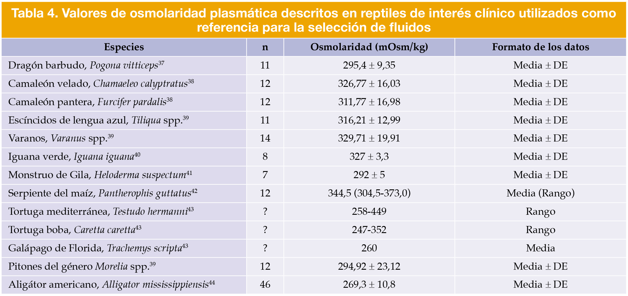
Mientras que los protocolos desactualizados utilizaban soluciones hipotónicas basadas en anfibios,[Martinez-Jimenez D, Hernandez-Divers SJ: Emergency care of reptiles. Vet Clin North Am Exot Anim Pract 2007; 10(2):557-585. [PubMed], Parkinson L: Fluid therapy in exotic animal emergency and critical care. Vet Clin North Am Exot Anim Pract 2023; 26(3):623-645. [PubMed], Kanda I, Brandão J: Nutrition and fluid therapy. En Graham JE, Beaufrère H, Doss GA (eds): Exotic Animal Emergency and Critical Care Medicine, 1ª ed., Hoboken, Wiley-Blackwell, 2021; 758-770.] los datos actuales indican que la osmolaridad plasmática de los reptiles oscila entre 260 y 370 mOsm/kg (Tabla 4).[Petritz OA, Son TT: Emergency and critical care. En Divers SJ, Stahl SJ (eds): Mader’s Reptile and Amphibian Medicine and Surgery, 3ª ed., St. Louis, Elsevier, 2019; 967-976., Parkinson L: Fluid therapy in exotic animal emergency and critical care. Vet Clin North Am Exot Anim Pract 2023; 26(3):623-645. [PubMed], Kanda I, Brandão J: Nutrition and fluid therapy. En Graham JE, Beaufrère H, Doss GA (eds): Exotic Animal Emergency and Critical Care Medicine, 1ª ed., Hoboken, Wiley-Blackwell, 2021; 758-770., Dallwig RK, Mitchell MA, Acierno MJ: Determination of plasma osmolality and agreement between measured and calculated values in healthy adult bearded dragons. J Herp Med Surg 2010; 20(2-3):69-73., Perry SM, Acierno MJ, Mitchell MA: Measuring the level of agreement between osmometer and calculated plasma osmolalities in two species of chameleons, Furcifer pardalis and Chamaeleo calyptratus. J Herp Med Surg 2021; 31(1):36-42., Cameron L, Mills B, Peters A: Plasma osmolality of Australian reptiles: are we assuming too much?. J Herp Med Surg 2024; 34(3):193-198., Fitzsimons JT, Kaufman S: Cellular and extracellular dehydration, and angiotensin as a stimuli to drinking in the common iguana (Iguana iguana). J Physiol 1977; 265(2):443-463. [PubMed], Davis JR, DeNardo DF: The urinary bladder as a physiologic reservoir that moderates dehydration in a large desert lizard, the Gila monster Heloderma suspectum. J Exp Biol 2007; 210:1472-1480. [PubMed], Sanchez-Migallon D, Mitchell MA, Acierno MJ: Determination of plasma osmolality and agreement between measured and calculated values in captive male corn snakes (Pantherophis [Elaphe] guttatus guttatus). J Herp Med Surg 2011; 21(1):16-19., Wilkinson R: Therapeutics. En McArthur S, Wilkinson R, Meyer J (eds): Medicine and Surgery of Tortoises and Turtles, Oxford, Blackwell Publishing, 2004; 465-485., Nevarez JG, Acierno MJ, Angel M, Beaufrere H: Determination of agreement between measured and calculated plasma osmolality values in captive-reared American alligators (Alligator mississippiensis). J Herp Med Surg 2012; 22(1-2):36-41.] En consecuencia, hoy en día se recomiendan cristaloides isotónicos, como NaCl 0,9 %, Plasma-Lyte A, Isofundin o Normosol-R.[Petritz OA, Son TT: Emergency and critical care. En Divers SJ, Stahl SJ (eds): Mader’s Reptile and Amphibian Medicine and Surgery, 3ª ed., St. Louis, Elsevier, 2019; 967-976., Music MK, Strunk A: Reptile critical care and common emergencies. Vet Clin North Am Exot Anim Pract 2016; 19(2):591-612. [PubMed], Parkinson L: Fluid therapy in exotic animal emergency and critical care. Vet Clin North Am Exot Anim Pract 2023; 26(3):623-645. [PubMed], Kanda I, Brandão J: Nutrition and fluid therapy. En Graham JE, Beaufrère H, Doss GA (eds): Exotic Animal Emergency and Critical Care Medicine, 1ª ed., Hoboken, Wiley-Blackwell, 2021; 758-770.] Además, el uso de soluciones con lactato puede emplearse de forma segura en animales con una función hepática normal.[Petritz OA, Son TT: Emergency and critical care. En Divers SJ, Stahl SJ (eds): Mader’s Reptile and Amphibian Medicine and Surgery, 3ª ed., St. Louis, Elsevier, 2019; 967-976., Music MK, Strunk A: Reptile critical care and common emergencies. Vet Clin North Am Exot Anim Pract 2016; 19(2):591-612. [PubMed], Martinez-Jimenez D, Hernandez-Divers SJ: Emergency care of reptiles. Vet Clin North Am Exot Anim Pract 2007; 10(2):557-585. [PubMed], Parkinson L: Fluid therapy in exotic animal emergency and critical care. Vet Clin North Am Exot Anim Pract 2023; 26(3):623-645. [PubMed], Kanda I, Brandão J: Nutrition and fluid therapy. En Graham JE, Beaufrère H, Doss GA (eds): Exotic Animal Emergency and Critical Care Medicine, 1ª ed., Hoboken, Wiley-Blackwell, 2021; 758-770.]
Tabla 4
Los reptiles metabolizan la dextrosa lentamente, por lo que los fluidos con glucosa pueden provocar hiperglucemia prolongada y agravar la deshidratación y recuperación del animal.[Parkinson L: Fluid therapy in exotic animal emergency and critical care. Vet Clin North Am Exot Anim Pract 2023; 26(3):623-645. [PubMed], Kanda I, Brandão J: Nutrition and fluid therapy. En Graham JE, Beaufrère H, Doss GA (eds): Exotic Animal Emergency and Critical Care Medicine, 1ª ed., Hoboken, Wiley-Blackwell, 2021; 758-770., Camacho M, Quintana MP, Calabuig P, et al: Acid-base and plasma biochemical changes using crystalloid fluids in stranded juvenile loggerhead sea turtles (Caretta caretta). PLoS One 2015; 10(7):e0132217. [PubMed], Parkinson L, Mans C: Evaluation of subcutaneously administered electrolyte solutions in experimentally dehydrated inland bearded dragons (Pogona vitticeps). Am J Vet Res 2020; 81(5):437-441. [PubMed]] En cuanto a los coloides, su uso debe evaluarse con cautela. Los coloides sintéticos se desaconsejan, ya que los reptiles mantienen su volumen intravascular mediante movilización intersticial, y su uso puede alterar la solubilidad del ácido úrico.[Petritz OA, Son TT: Emergency and critical care. En Divers SJ, Stahl SJ (eds): Mader’s Reptile and Amphibian Medicine and Surgery, 3ª ed., St. Louis, Elsevier, 2019; 967-976., Parkinson L: Fluid therapy in exotic animal emergency and critical care. Vet Clin North Am Exot Anim Pract 2023; 26(3):623-645. [PubMed], Braun EJ: Integration of organ systems in avian osmoregulation. J Exp Zool 1999; 283(7):702-707.] En casos de hipoalbuminemia, pueden considerarse los coloides naturales, como el plasma o la albúmina de reptil, para restaurar la presión oncótica y prevenir ascitis.[Petritz OA, Son TT: Emergency and critical care. En Divers SJ, Stahl SJ (eds): Mader’s Reptile and Amphibian Medicine and Surgery, 3ª ed., St. Louis, Elsevier, 2019; 967-976., Parkinson L: Fluid therapy in exotic animal emergency and critical care. Vet Clin North Am Exot Anim Pract 2023; 26(3):623-645. [PubMed]]
Velocidad de administración
Los requerimientos de mantenimiento son de 5-25 ml/kg/día, con el objetivo de una rehidratación completa en 48-96 horas.[Wilkinson SL: Physical examination, triage, and stabilization of the critical reptile patient. LafeberVet Webinar Library: The Critical Reptile Patient. Lafeber Company, 2023. Disponible en: https://lafeber.com/vet/the-critical-reptile-patient/, Petritz OA, Son TT: Emergency and critical care. En Divers SJ, Stahl SJ (eds): Mader’s Reptile and Amphibian Medicine and Surgery, 3ª ed., St. Louis, Elsevier, 2019; 967-976., Martinez-Jimenez D, Hernandez-Divers SJ: Emergency care of reptiles. Vet Clin North Am Exot Anim Pract 2007; 10(2):557-585. [PubMed], Kanda I, Brandão J: Nutrition and fluid therapy. En Graham JE, Beaufrère H, Doss GA (eds): Exotic Animal Emergency and Critical Care Medicine, 1ª ed., Hoboken, Wiley-Blackwell, 2021; 758-770.] No obstante, estos requerimientos deben elegirse en función de la especie y su ecosistema de origen, siendo generalmente menores en reptiles adaptados a ambientes áridos y mayores en especies tropicales o acuáticas. Los fluidos deben atemperarse hasta la POTZ antes de su administración, ya que los reptiles hipotérmicos presentan una absorción deficiente.[Wilkinson SL: Physical examination, triage, and stabilization of the critical reptile patient. LafeberVet Webinar Library: The Critical Reptile Patient. Lafeber Company, 2023. Disponible en: https://lafeber.com/vet/the-critical-reptile-patient/, Petritz OA, Son TT: Emergency and critical care. En Divers SJ, Stahl SJ (eds): Mader’s Reptile and Amphibian Medicine and Surgery, 3ª ed., St. Louis, Elsevier, 2019; 967-976., Martinez-Jimenez D, Hernandez-Divers SJ: Emergency care of reptiles. Vet Clin North Am Exot Anim Pract 2007; 10(2):557-585. [PubMed]]
Transfusión sanguínea
Aunque muchos reptiles toleran grados extremos de anemia, e incluso algunas especies pueden mantenerse estables con un hematocrito (PCV) cercano al 5 % siempre que se encuentren normotérmicos e hidratados, la transfusión sanguínea puede resultar beneficiosa en casos de hemorragia grave, PCV <10–12 % o hipoproteinemia marcada.[Parkinson L: Fluid therapy in exotic animal emergency and critical care. Vet Clin North Am Exot Anim Pract 2023; 26(3):623-645. [PubMed], Kanda I, Brandão J: Nutrition and fluid therapy. En Graham JE, Beaufrère H, Doss GA (eds): Exotic Animal Emergency and Critical Care Medicine, 1ª ed., Hoboken, Wiley-Blackwell, 2021; 758-770.]
Dado que no existen pruebas de compatibilidad (crossmatch) para reptiles, los donantes deben ser congéneres clínicamente sanos, libres de enfermedades y de tamaño similar.[Petritz OA, Son TT: Emergency and critical care. En Divers SJ, Stahl SJ (eds): Mader’s Reptile and Amphibian Medicine and Surgery, 3ª ed., St. Louis, Elsevier, 2019; 967-976., Kanda I, Brandão J: Nutrition and fluid therapy. En Graham JE, Beaufrère H, Doss GA (eds): Exotic Animal Emergency and Critical Care Medicine, 1ª ed., Hoboken, Wiley-Blackwell, 2021; 758-770.] En situaciones excepcionales, pueden intentarse transfusiones interespecíficas entre taxones estrechamente relacionados, aunque con la debida precaución.[Petritz OA, Son TT: Emergency and critical care. En Divers SJ, Stahl SJ (eds): Mader’s Reptile and Amphibian Medicine and Surgery, 3ª ed., St. Louis, Elsevier, 2019; 967-976., Kanda I, Brandão J: Nutrition and fluid therapy. En Graham JE, Beaufrère H, Doss GA (eds): Exotic Animal Emergency and Critical Care Medicine, 1ª ed., Hoboken, Wiley-Blackwell, 2021; 758-770.]
Se debe extraer un volumen máximo de 5-10 ml/kg, utilizando CPDA (citrato, fosfato, dextrosa y adenina) en proporción 1:9 o heparina (5-10 UI/ml) como anticoagulante.[Petritz OA, Son TT: Emergency and critical care. En Divers SJ, Stahl SJ (eds): Mader’s Reptile and Amphibian Medicine and Surgery, 3ª ed., St. Louis, Elsevier, 2019; 967-976., Schumacher J: Fluid therapy in reptiles. En Kirk RW (ed): Current Veterinary Therapy XIII. Philadelphia, WB Saunders, 2000; 1170-1173.] Las muestras deben mantenerse refrigeradas y utilizarse en un plazo máximo de 6 horas. La administración se realiza por vía intravenosa (IV) o intraósea (IO), empleando un filtro para sangre de 18 µm.[Petritz OA, Son TT: Emergency and critical care. En Divers SJ, Stahl SJ (eds): Mader’s Reptile and Amphibian Medicine and Surgery, 3ª ed., St. Louis, Elsevier, 2019; 967-976., Kanda I, Brandão J: Nutrition and fluid therapy. En Graham JE, Beaufrère H, Doss GA (eds): Exotic Animal Emergency and Critical Care Medicine, 1ª ed., Hoboken, Wiley-Blackwell, 2021; 758-770., Nevarez JG, Cockburn J, Kearney MT, et al: Evaluation of an 18-micron filter for use in reptile blood transfusions using blood from American alligators (Alligator mississippiensis). J Zoo Wildl Med 2011; 42:236-240. [PubMed]] La sangre debe calentarse a 25-30 °C antes de la transfusión.[Petritz OA, Son TT: Emergency and critical care. En Divers SJ, Stahl SJ (eds): Mader’s Reptile and Amphibian Medicine and Surgery, 3ª ed., St. Louis, Elsevier, 2019; 967-976., Schumacher J: Fluid therapy in reptiles. En Kirk RW (ed): Current Veterinary Therapy XIII. Philadelphia, WB Saunders, 2000; 1170-1173.]
La dosis recomendada es de 10-15 ml/kg, iniciando la transfusión lentamente (0,5-1 ml/kg durante los primeros 10-15 minutos) para detectar posibles reacciones adversas, y aumentando posteriormente la velocidad de infusión de forma gradual mientras se monitorizan la frecuencia cardíaca, el color de las mucosas y la perfusión periférica.[Petritz OA, Son TT: Emergency and critical care. En Divers SJ, Stahl SJ (eds): Mader’s Reptile and Amphibian Medicine and Surgery, 3ª ed., St. Louis, Elsevier, 2019; 967-976., Schumacher J: Fluid therapy in reptiles. En Kirk RW (ed): Current Veterinary Therapy XIII. Philadelphia, WB Saunders, 2000; 1170-1173.] Se debe revaluar el PCV y las proteínas totales 1-2 horas después; un aumento del 5-10 % en el PCV indica una reposición adecuada.
Analgesia y terapias complementarias
Analgesia
La evidencia sobre el manejo del dolor en reptiles sigue siendo limitada y la mayoría de los protocolos se basan en extrapolaciones de otras especies.[Long SY: Approach to reptile emergency medicine. Vet Clin North Am Exot Anim Pract 2016; 19(2):567-590. [PubMed], Sladky KK, Mans C: Analgesia. En Divers SJ, Stahl SJ (eds): Mader’s Reptile and Amphibian Medicine and Surgery, 3ª ed., St. Louis, Elsevier, 2019; 465-474.] La analgesia local, con lidocaína (2-4 mg/kg) o bupivacaína (1 mg/kg), puede combinarse con morfina (0,1-0,2 mg/kg) para su administración por vía intratecal, proporcionando 24-48 horas de analgesia efectiva.[Wilkinson SL: Physical examination, triage, and stabilization of the critical reptile patient. LafeberVet Webinar Library: The Critical Reptile Patient. Lafeber Company, 2023. Disponible en: https://lafeber.com/vet/the-critical-reptile-patient/, Martinez-Jimenez D, Hernandez-Divers SJ: Emergency care of reptiles. Vet Clin North Am Exot Anim Pract 2007; 10(2):557-585. [PubMed], Sladky KK, Klaphake E, Di Girolamo N, Carpenter JW: Reptiles. En Carpenter JW, Marion CJ (eds): Carpenter’s Exotic Animal Formulary, 6ª ed., St. Louis, Elsevier, 2023; 101-173., Sladky KK, Mans C: Analgesia. En Divers SJ, Stahl SJ (eds): Mader’s Reptile and Amphibian Medicine and Surgery, 3ª ed., St. Louis, Elsevier, 2019; 465-474., Doss GA, Mans C, Sladky KK: Analgesia, anesthesia, and monitoring. En Graham JE, Beaufrère H, Doss GA (eds): Exotic Animal Emergency and Critical Care Medicine, 1ª ed., Hoboken, Wiley-Blackwell, 2021; 746-757., Sladky KK, Mans C: Clinical analgesia in reptiles. J Exot Pet Med 2012; 21(2):158-167.] Esta técnica se ha descrito principalmente en quelonios, así como en algunos saurios de mayor tamaño como iguanas, tegus y dragones barbudos; no obstante, en quelonios existen diferencias anatómicas relevantes, como la presencia de un plexo venoso intravertebral prominente, que pueden dificultar la estandarización de la técnica entre especies.[Mans C, Steagall P, Sladky KK: Regional Anesthesia and Analgesia. En Divers SJ, Stahl SJ (eds): Mader’s Reptile and Amphibian Medicine and Surgery, 3ª ed., St. Louis, Elsevier, 2019; 475-480.]
La eficacia analgésica de los antiinflamatorios no esteroideos (AINEs) no ha sido demostrada en estudios controlados: la administración PO presenta una absorción deficiente y niveles plasmáticos subterapéuticos, mientras que la vía intramuscular ofrece concentraciones más estables.[Martinez-Jimenez D, Hernandez-Divers SJ: Emergency care of reptiles. Vet Clin North Am Exot Anim Pract 2007; 10(2):557-585. [PubMed], Sladky KK, Klaphake E, Di Girolamo N, Carpenter JW: Reptiles. En Carpenter JW, Marion CJ (eds): Carpenter’s Exotic Animal Formulary, 6ª ed., St. Louis, Elsevier, 2023; 101-173., Sladky KK, Mans C: Analgesia. En Divers SJ, Stahl SJ (eds): Mader’s Reptile and Amphibian Medicine and Surgery, 3ª ed., St. Louis, Elsevier, 2019; 465-474., Doss GA, Mans C, Sladky KK: Analgesia, anesthesia, and monitoring. En Graham JE, Beaufrère H, Doss GA (eds): Exotic Animal Emergency and Critical Care Medicine, 1ª ed., Hoboken, Wiley-Blackwell, 2021; 746-757., Sladky KK, Mans C: Clinical analgesia in reptiles. J Exot Pet Med 2012; 21(2):158-167.] Se ha sugerido que la COX-1 podría tener un papel predominante en la inflamación en reptiles, lo que apunta a que el ketoprofeno (2 mg/kg IM cada 24 h) podría ser una alternativa más precisa al meloxicam (inhibidor selectivo de COX-2).[Wilkinson SL: Physical examination, triage, and stabilization of the critical reptile patient. LafeberVet Webinar Library: The Critical Reptile Patient. Lafeber Company, 2023. Disponible en: https://lafeber.com/vet/the-critical-reptile-patient/, Sladky KK, Klaphake E, Di Girolamo N, Carpenter JW: Reptiles. En Carpenter JW, Marion CJ (eds): Carpenter’s Exotic Animal Formulary, 6ª ed., St. Louis, Elsevier, 2023; 101-173., Sladky KK, Mans C: Analgesia. En Divers SJ, Stahl SJ (eds): Mader’s Reptile and Amphibian Medicine and Surgery, 3ª ed., St. Louis, Elsevier, 2019; 465-474., Sladky KK, Mans C: Clinical analgesia in reptiles. J Exot Pet Med 2012; 21(2):158-167.]
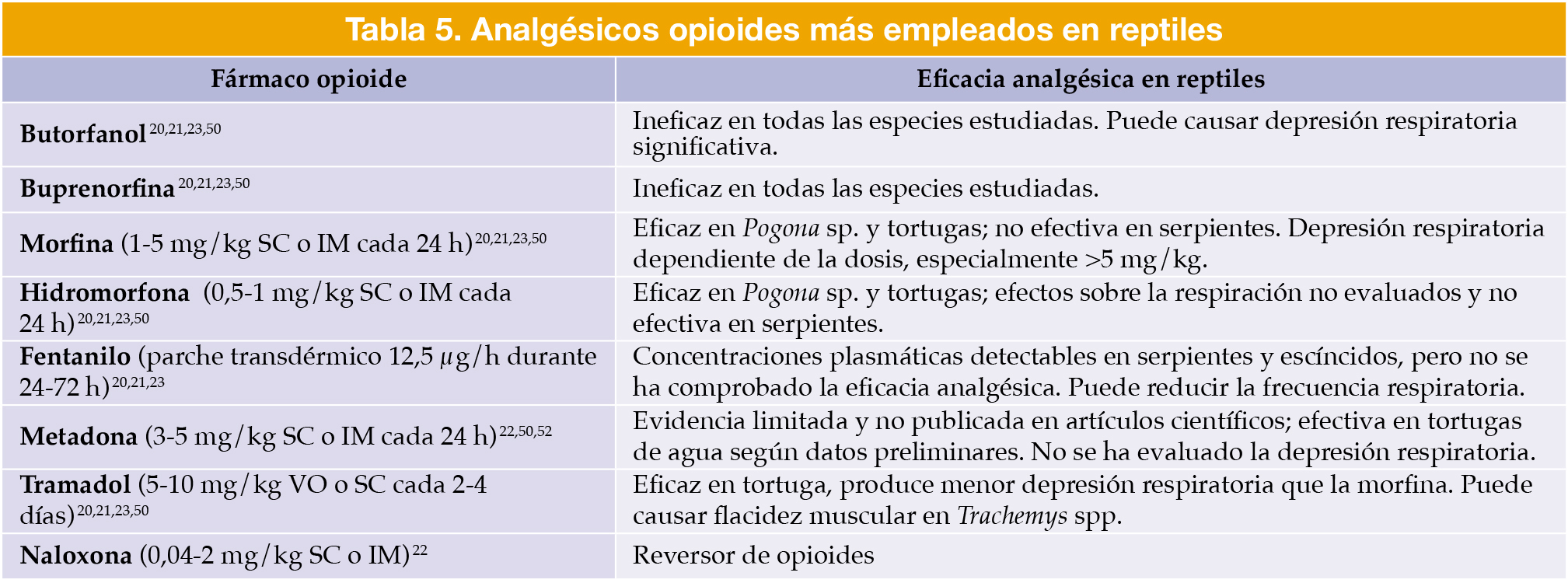
Los reptiles expresan receptores opioides de los subtipos μ (mu) y predominantemente δ (delta).[Doss GA, Mans C, Sladky KK: Analgesia, anesthesia, and monitoring. En Graham JE, Beaufrère H, Doss GA (eds): Exotic Animal Emergency and Critical Care Medicine, 1ª ed., Hoboken, Wiley-Blackwell, 2021; 746-757., Burggren WW, Christoffels VM, Crossley DA 2nd, et al: Comparative cardiovascular physiology: future trends, opportunities and challenges. Acta Physiol 2014; 210(2):257-276. [PubMed], Sladky KK, Mans C: Clinical analgesia in reptiles. J Exot Pet Med 2012; 21(2):158-167.] Los opioides más empleados y su eficacia en reptiles se resumen en la Tabla 5.[Sladky KK, Klaphake E, Di Girolamo N, Carpenter JW: Reptiles. En Carpenter JW, Marion CJ (eds): Carpenter’s Exotic Animal Formulary, 6ª ed., St. Louis, Elsevier, 2023; 101-173., Sladky KK, Mans C: Analgesia. En Divers SJ, Stahl SJ (eds): Mader’s Reptile and Amphibian Medicine and Surgery, 3ª ed., St. Louis, Elsevier, 2019; 465-474., Doss GA, Mans C, Sladky KK: Analgesia, anesthesia, and monitoring. En Graham JE, Beaufrère H, Doss GA (eds): Exotic Animal Emergency and Critical Care Medicine, 1ª ed., Hoboken, Wiley-Blackwell, 2021; 746-757., Sladky KK, Mans C: Clinical analgesia in reptiles. J Exot Pet Med 2012; 21(2):158-167., Sladky KK, Johnson SM: Current understanding of analgesic efficacy and associated side effects in reptiles. Proc Annu Conf Am Assoc Zoo Vet, Los Angeles, CA, 2008; 116-117.]
Tabla 5
SC: subcutáneo; IM: intramuscular; VO: vía oral.
Antibióticos y terapias complementarias
Los antibióticos rara vez son necesarios durante la fase inicial de urgencia, antes de que el animal esté estabilizado.[Martinez-Jimenez D, Hernandez-Divers SJ: Emergency care of reptiles. Vet Clin North Am Exot Anim Pract 2007; 10(2):557-585. [PubMed]] El tratamiento debe iniciarse únicamente cuando el reptil se encuentre normotérmico e hidratado, ya que la absorción y el metabolismo de los fármacos dependen de la temperatura corporal.[Petritz OA, Son TT: Emergency and critical care. En Divers SJ, Stahl SJ (eds): Mader’s Reptile and Amphibian Medicine and Surgery, 3ª ed., St. Louis, Elsevier, 2019; 967-976., Long SY: Approach to reptile emergency medicine. Vet Clin North Am Exot Anim Pract 2016; 19(2):567-590. [PubMed], Martinez-Jimenez D, Hernandez-Divers SJ: Emergency care of reptiles. Vet Clin North Am Exot Anim Pract 2007; 10(2):557-585. [PubMed]] Aunque no se recomienda, si se inicia un tratamiento antibiótico mientras se esperan los resultados del cultivo, este debe dirigirse principalmente contra bacterias gramnegativas.[Wilkinson SL: Physical examination, triage, and stabilization of the critical reptile patient. LafeberVet Webinar Library: The Critical Reptile Patient. Lafeber Company, 2023. Disponible en: https://lafeber.com/vet/the-critical-reptile-patient/, Petritz OA, Son TT: Emergency and critical care. En Divers SJ, Stahl SJ (eds): Mader’s Reptile and Amphibian Medicine and Surgery, 3ª ed., St. Louis, Elsevier, 2019; 967-976., Martinez-Jimenez D, Hernandez-Divers SJ: Emergency care of reptiles. Vet Clin North Am Exot Anim Pract 2007; 10(2):557-585. [PubMed]]
Con respecto a las terapias complementarias, puede administrarse gluconato cálcico (100 mg/kg SC, IM, o IV cada 6-24 h, y cambiar a PO una vez estabilizado) en casos de temblores musculares por hipocalcemia, y alopurinol (10-50 mg/kg (normalmente 25 mg/kg) PO cada 24 h) en presencia de hiperuricemia, según indicación clínica.[Petritz OA, Son TT: Emergency and critical care. En Divers SJ, Stahl SJ (eds): Mader’s Reptile and Amphibian Medicine and Surgery, 3ª ed., St. Louis, Elsevier, 2019; 967-976., Martinez-Jimenez D, Hernandez-Divers SJ: Emergency care of reptiles. Vet Clin North Am Exot Anim Pract 2007; 10(2):557-585. [PubMed], Sladky KK, Klaphake E, Di Girolamo N, Carpenter JW: Reptiles. En Carpenter JW, Marion CJ (eds): Carpenter’s Exotic Animal Formulary, 6ª ed., St. Louis, Elsevier, 2023; 101-173.] De forma adicional, se ha descrito el uso de rasburicasa (0,2 mg/kg IM) como alternativa uricolítica en reptiles.[Cope I: Fasturtec: A new treatment for renal disease in reptiles and birds?. Proc Int Conf Avian Herpetol Exotic Mamm Med, Wiesbaden, Germany, 2013; 331., Montesinos A, Ardiaca M, Bonvehí LV, et al: Effects of treatment with one dose of rasburicase on plasma uric acid levels of red-eared slider (Trachemys scripta) and Greek tortoises (Testudo graeca). Proc Int Conf Avian Herpetol Exotic Mamm Med, Budapest, Hungary, 2022.] Aunque su utilización se ha extendido en algunos entornos clínicos, los datos disponibles proceden únicamente de dos comunicaciones científicas preliminares, por lo que la evidencia actual resulta insuficiente para establecer protocolos de administración estandarizados en la literatura.
Soporte nutricional
La nutrición debe iniciarse una vez que el paciente esté estabilizado.[Wilkinson SL: Physical examination, triage, and stabilization of the critical reptile patient. LafeberVet Webinar Library: The Critical Reptile Patient. Lafeber Company, 2023. Disponible en: https://lafeber.com/vet/the-critical-reptile-patient/, Long SY: Approach to reptile emergency medicine. Vet Clin North Am Exot Anim Pract 2016; 19(2):567-590. [PubMed]] Los requerimientos energéticos pueden estimarse mediante la fórmula:[Martinez-Jimenez D, Hernandez-Divers SJ: Emergency care of reptiles. Vet Clin North Am Exot Anim Pract 2007; 10(2):557-585. [PubMed], Kanda I, Brandão J: Nutrition and fluid therapy. En Graham JE, Beaufrère H, Doss GA (eds): Exotic Animal Emergency and Critical Care Medicine, 1ª ed., Hoboken, Wiley-Blackwell, 2021; 758-770.]
Posteriormente, deben aplicarse factores de corrección según el estado fisiológico del animal para obtener la tasa metabólica de mantenimiento (MMR, kcal/día):[Martinez-Jimenez D, Hernandez-Divers SJ: Emergency care of reptiles. Vet Clin North Am Exot Anim Pract 2007; 10(2):557-585. [PubMed], Kanda I, Brandão J: Nutrition and fluid therapy. En Graham JE, Beaufrère H, Doss GA (eds): Exotic Animal Emergency and Critical Care Medicine, 1ª ed., Hoboken, Wiley-Blackwell, 2021; 758-770.]
Ayuno o inanición: × 0,25-0,75
Actividad o cirugía: × 1-1,25
Traumatismo o sepsis: × 1-1,5
Cicatrización o reproducción: × 1,5
Estrés o crecimiento: × 2
Para calcular el volumen total de la fórmula nutricional y el esquema de alimentación, se utiliza la relación:[Martinez-Jimenez D, Hernandez-Divers SJ: Emergency care of reptiles. Vet Clin North Am Exot Anim Pract 2007; 10(2):557-585. [PubMed], Kanda I, Brandão J: Nutrition and fluid therapy. En Graham JE, Beaufrère H, Doss GA (eds): Exotic Animal Emergency and Critical Care Medicine, 1ª ed., Hoboken, Wiley-Blackwell, 2021; 758-770.]
MMR ÷ kcal/ml (de la fórmula nutricional seleccionada) = ml de fórmula por día
En pacientes con anorexia prolongada o desnutrición, la realimentación debe iniciarse de forma gradual, comenzando con aproximadamente el 10-25 % de los requerimientos calculados y aumentando progresivamente durante varios días o semanas.[Martinez-Jimenez D, Hernandez-Divers SJ: Emergency care of reptiles. Vet Clin North Am Exot Anim Pract 2007; 10(2):557-585. [PubMed], Kanda I, Brandão J: Nutrition and fluid therapy. En Graham JE, Beaufrère H, Doss GA (eds): Exotic Animal Emergency and Critical Care Medicine, 1ª ed., Hoboken, Wiley-Blackwell, 2021; 758-770.] Aunque el síndrome de realimentación está bien descrito en mamíferos, su frecuencia en reptiles no está claramente establecida; no obstante, se recomienda monitorizar estrechamente electrolitos como fósforo, potasio y magnesio, así como la glucemia, especialmente durante los primeros días de soporte nutricional.[Martinez-Jimenez D, Hernandez-Divers SJ: Emergency care of reptiles. Vet Clin North Am Exot Anim Pract 2007; 10(2):557-585. [PubMed], Kanda I, Brandão J: Nutrition and fluid therapy. En Graham JE, Beaufrère H, Doss GA (eds): Exotic Animal Emergency and Critical Care Medicine, 1ª ed., Hoboken, Wiley-Blackwell, 2021; 758-770.]
Pruebas diagnósticas
Las pruebas diagnósticas en una urgencia deben realizarse tras la estabilización y la normalización de la temperatura corporal, ya que la hipotermia puede alterar los valores analíticos.[Petritz OA, Son TT: Emergency and critical care. En Divers SJ, Stahl SJ (eds): Mader’s Reptile and Amphibian Medicine and Surgery, 3ª ed., St. Louis, Elsevier, 2019; 967-976., Long SY: Approach to reptile emergency medicine. Vet Clin North Am Exot Anim Pract 2016; 19(2):567-590. [PubMed], Martinez-Jimenez D, Hernandez-Divers SJ: Emergency care of reptiles. Vet Clin North Am Exot Anim Pract 2007; 10(2):557-585. [PubMed]] Los diferentes métodos que ayudan a orientar el diagnóstico y el tratamiento en pacientes reptiles son, en gran medida, similares a los empleados en mamíferos y aves. Debido al enfoque práctico del presente artículo y a las limitaciones de extensión editorial, las herramientas diagnósticas no se describen de forma exhaustiva. Para una revisión más detallada de estas técnicas en medicina de reptiles, se recomienda consultar la literatura especializada. Estos procedimientos diagnósticos incluyen los presentados a continuación.
Analíticas sanguíneas
El hemograma completo (CBC), la determinación del hematocrito o microhematocrito (PCV) y la determinación de proteínas totales mediante refractometría constituyen herramientas rápidas especialmente útiles en el paciente crítico para la detección precoz de deshidratación, anemia y cambios en los glóbulos blancos indicativos de inflamación o infección sistémica.[Petritz OA, Son TT: Emergency and critical care. En Divers SJ, Stahl SJ (eds): Mader’s Reptile and Amphibian Medicine and Surgery, 3ª ed., St. Louis, Elsevier, 2019; 967-976., Martinez-Jimenez D, Hernandez-Divers SJ: Emergency care of reptiles. Vet Clin North Am Exot Anim Pract 2007; 10(2):557-585. [PubMed], DiGeronimo PM, Di Girolamo N: STAT diagnostics. En Graham JE, Beaufrère H, Doss GA (eds): Exotic Animal Emergency and Critical Care Medicine, 1ª ed., Hoboken, Wiley-Blackwell, 2021; 771-782.] Dado que los eritrocitos de los reptiles son nucleados, la evaluación manual de los frotis sanguíneos sigue siendo esencial.[Music MK, Strunk A: Reptile critical care and common emergencies. Vet Clin North Am Exot Anim Pract 2016; 19(2):591-612. [PubMed], Doss GA, Mans C, Sladky KK: Analgesia, anesthesia, and monitoring. En Graham JE, Beaufrère H, Doss GA (eds): Exotic Animal Emergency and Critical Care Medicine, 1ª ed., Hoboken, Wiley-Blackwell, 2021; 746-757.]
Los perfiles bioquímicos permiten valorar la funcionalidad de ciertos órganos y el equilibrio electrolítico; los analizadores bioquímicos veterinarios de pequeño volumen (p. ej., VetScan 2®, Zoetis) resultan especialmente útiles en especies de tamaño reducido.[Wilkinson SL: Physical examination, triage, and stabilization of the critical reptile patient. LafeberVet Webinar Library: The Critical Reptile Patient. Lafeber Company, 2023. Disponible en: https://lafeber.com/vet/the-critical-reptile-patient/, Petritz OA, Son TT: Emergency and critical care. En Divers SJ, Stahl SJ (eds): Mader’s Reptile and Amphibian Medicine and Surgery, 3ª ed., St. Louis, Elsevier, 2019; 967-976., Music MK, Strunk A: Reptile critical care and common emergencies. Vet Clin North Am Exot Anim Pract 2016; 19(2):591-612. [PubMed], Martinez-Jimenez D, Hernandez-Divers SJ: Emergency care of reptiles. Vet Clin North Am Exot Anim Pract 2007; 10(2):557-585. [PubMed], DiGeronimo PM, Di Girolamo N: STAT diagnostics. En Graham JE, Beaufrère H, Doss GA (eds): Exotic Animal Emergency and Critical Care Medicine, 1ª ed., Hoboken, Wiley-Blackwell, 2021; 771-782.] Mientras que el ácido úrico es el principal producto nitrogenado en reptiles terrestres, en las tortugas de agua dulce suele predominar la urea y en cocodrilos y tortugas marinas, el amoníaco.[Petritz OA, Son TT: Emergency and critical care. En Divers SJ, Stahl SJ (eds): Mader’s Reptile and Amphibian Medicine and Surgery, 3ª ed., St. Louis, Elsevier, 2019; 967-976., Martinez-Jimenez D, Hernandez-Divers SJ: Emergency care of reptiles. Vet Clin North Am Exot Anim Pract 2007; 10(2):557-585. [PubMed], DiGeronimo PM, Di Girolamo N: STAT diagnostics. En Graham JE, Beaufrère H, Doss GA (eds): Exotic Animal Emergency and Critical Care Medicine, 1ª ed., Hoboken, Wiley-Blackwell, 2021; 771-782.]
Los analizadores bioquímicos y de gases portátiles (p. ej., i-STAT®, Zoetis) permiten obtener resultados rápidos y precisos, principalmente para la medición de gases sanguíneos, calcio ionizado y electrolitos.[Wilkinson SL: Physical examination, triage, and stabilization of the critical reptile patient. LafeberVet Webinar Library: The Critical Reptile Patient. Lafeber Company, 2023. Disponible en: https://lafeber.com/vet/the-critical-reptile-patient/, Music MK, Strunk A: Reptile critical care and common emergencies. Vet Clin North Am Exot Anim Pract 2016; 19(2):591-612. [PubMed], Martinez-Jimenez D, Hernandez-Divers SJ: Emergency care of reptiles. Vet Clin North Am Exot Anim Pract 2007; 10(2):557-585. [PubMed], DiGeronimo PM, Di Girolamo N: STAT diagnostics. En Graham JE, Beaufrère H, Doss GA (eds): Exotic Animal Emergency and Critical Care Medicine, 1ª ed., Hoboken, Wiley-Blackwell, 2021; 771-782.] Es necesario recordar que los gases sanguíneos deben analizarse en la POTZ del animal.[Petritz OA, Son TT: Emergency and critical care. En Divers SJ, Stahl SJ (eds): Mader’s Reptile and Amphibian Medicine and Surgery, 3ª ed., St. Louis, Elsevier, 2019; 967-976., Martinez-Jimenez D, Hernandez-Divers SJ: Emergency care of reptiles. Vet Clin North Am Exot Anim Pract 2007; 10(2):557-585. [PubMed], DiGeronimo PM, Di Girolamo N: STAT diagnostics. En Graham JE, Beaufrère H, Doss GA (eds): Exotic Animal Emergency and Critical Care Medicine, 1ª ed., Hoboken, Wiley-Blackwell, 2021; 771-782.] Los valores normales de pH oscilan entre 7,5 y 7,7, con un CO₂ total de 20-30 mmol/l.[Petritz OA, Son TT: Emergency and critical care. En Divers SJ, Stahl SJ (eds): Mader’s Reptile and Amphibian Medicine and Surgery, 3ª ed., St. Louis, Elsevier, 2019; 967-976., Martinez-Jimenez D, Hernandez-Divers SJ: Emergency care of reptiles. Vet Clin North Am Exot Anim Pract 2007; 10(2):557-585. [PubMed], DiGeronimo PM, Di Girolamo N: STAT diagnostics. En Graham JE, Beaufrère H, Doss GA (eds): Exotic Animal Emergency and Critical Care Medicine, 1ª ed., Hoboken, Wiley-Blackwell, 2021; 771-782.] La medición del calcio ionizado es especialmente útil en hembras reproductoras o en pacientes con enfermedad ósea metabólica.[Wilkinson SL: Physical examination, triage, and stabilization of the critical reptile patient. LafeberVet Webinar Library: The Critical Reptile Patient. Lafeber Company, 2023. Disponible en: https://lafeber.com/vet/the-critical-reptile-patient/, Petritz OA, Son TT: Emergency and critical care. En Divers SJ, Stahl SJ (eds): Mader’s Reptile and Amphibian Medicine and Surgery, 3ª ed., St. Louis, Elsevier, 2019; 967-976., Music MK, Strunk A: Reptile critical care and common emergencies. Vet Clin North Am Exot Anim Pract 2016; 19(2):591-612. [PubMed], Martinez-Jimenez D, Hernandez-Divers SJ: Emergency care of reptiles. Vet Clin North Am Exot Anim Pract 2007; 10(2):557-585. [PubMed], DiGeronimo PM, Di Girolamo N: STAT diagnostics. En Graham JE, Beaufrère H, Doss GA (eds): Exotic Animal Emergency and Critical Care Medicine, 1ª ed., Hoboken, Wiley-Blackwell, 2021; 771-782.] En tortugas, un valor de corte de 2,2 mmol/l en la diferencia entre calcio total y calcio ionizado (tCa - iCa) puede ayudar a diferenciar animales con o sin alteraciones del aparato reproductor.[Di Girolamo N: Relationship, difference, and diagnostic discordance between blood ionized and total calcium concentrations in client-owned chelonians. J Am Vet Med Assoc 2022; 260(S2):S101-S110. [PubMed]]
Diagnóstico por imagen
La ecografía POCUS (Point-of-Care Ultrasound) permite una evaluación rápida en tiempo real de los órganos celómicos en el paciente crítico, incluyendo la detección de líquido libre celómico, la valoración de la vejiga urinaria, la evaluación cardíaca básica y la identificación del estado reproductivo.[Petritz OA, Son TT: Emergency and critical care. En Divers SJ, Stahl SJ (eds): Mader’s Reptile and Amphibian Medicine and Surgery, 3ª ed., St. Louis, Elsevier, 2019; 967-976., Music MK, Strunk A: Reptile critical care and common emergencies. Vet Clin North Am Exot Anim Pract 2016; 19(2):591-612. [PubMed], Martinez-Jimenez D, Hernandez-Divers SJ: Emergency care of reptiles. Vet Clin North Am Exot Anim Pract 2007; 10(2):557-585. [PubMed], Gardhouse S: Point of care ultrasound in exotic animal emergency and critical care. Vet Clin Exot Anim Pract 2023; 26(3):567-589. [PubMed], Fazio C: Diagnostic imaging. En Graham JE, Beaufrère H, Doss GA (eds): Exotic Animal Emergency and Critical Care Medicine, 1ª ed., Hoboken, Wiley-Blackwell, 2021; 783-799.] La radiografía complementa la ecografía al permitir de forma más sencilla la detección de huevos, urolitos, enfermedad respiratoria, lesiones óseas y alteraciones digestivas como fecalomas, impactaciones u otros procesos obstructivos.[Wilkinson SL: Physical examination, triage, and stabilization of the critical reptile patient. LafeberVet Webinar Library: The Critical Reptile Patient. Lafeber Company, 2023. Disponible en: https://lafeber.com/vet/the-critical-reptile-patient/, Petritz OA, Son TT: Emergency and critical care. En Divers SJ, Stahl SJ (eds): Mader’s Reptile and Amphibian Medicine and Surgery, 3ª ed., St. Louis, Elsevier, 2019; 967-976., Music MK, Strunk A: Reptile critical care and common emergencies. Vet Clin North Am Exot Anim Pract 2016; 19(2):591-612. [PubMed], Martinez-Jimenez D, Hernandez-Divers SJ: Emergency care of reptiles. Vet Clin North Am Exot Anim Pract 2007; 10(2):557-585. [PubMed], Fazio C: Diagnostic imaging. En Graham JE, Beaufrère H, Doss GA (eds): Exotic Animal Emergency and Critical Care Medicine, 1ª ed., Hoboken, Wiley-Blackwell, 2021; 783-799.] Las técnicas avanzadas de imagen, como la tomografía computarizada o la resonancia magnética, ofrecen una visualización de alta resolución de los sistemas respiratorio o neurológico, pero deben reservarse para pacientes estables que toleren anestesia.[Wilkinson SL: Physical examination, triage, and stabilization of the critical reptile patient. LafeberVet Webinar Library: The Critical Reptile Patient. Lafeber Company, 2023. Disponible en: https://lafeber.com/vet/the-critical-reptile-patient/, Petritz OA, Son TT: Emergency and critical care. En Divers SJ, Stahl SJ (eds): Mader’s Reptile and Amphibian Medicine and Surgery, 3ª ed., St. Louis, Elsevier, 2019; 967-976., Fazio C: Diagnostic imaging. En Graham JE, Beaufrère H, Doss GA (eds): Exotic Animal Emergency and Critical Care Medicine, 1ª ed., Hoboken, Wiley-Blackwell, 2021; 783-799.]
La endoscopia constituye una herramienta diagnóstica mínimamente invasiva de gran utilidad en reptiles clínicamente estables, ya que permite la visualización directa de las vías digestivas, respiratorias y urogenitales, así como la obtención de muestras dirigidas para citología, cultivo o biopsia.[Divers SJ: Reptile diagnostic endoscopy and endosurgery. Vet Clin North Am Exot Anim Pract 2010;13(2):217-42. [PubMed]] En muchos casos facilita el diagnóstico definitivo y puede evitar procedimientos más invasivos como la celioscopia o la celotomía exploratoria.
Otras pruebas diagnósticas
El análisis coprológico es recomendable, dado que el parasitismo constituye una causa frecuente de enfermedad sistémica.[Wilkinson SL: Physical examination, triage, and stabilization of the critical reptile patient. LafeberVet Webinar Library: The Critical Reptile Patient. Lafeber Company, 2023. Disponible en: https://lafeber.com/vet/the-critical-reptile-patient/, DiGeronimo PM, Di Girolamo N: STAT diagnostics. En Graham JE, Beaufrère H, Doss GA (eds): Exotic Animal Emergency and Critical Care Medicine, 1ª ed., Hoboken, Wiley-Blackwell, 2021; 771-782.]
Los cultivos y pruebas de sensibilidad antibiótica orientan la selección del tratamiento antimicrobiano;[Wilkinson SL: Physical examination, triage, and stabilization of the critical reptile patient. LafeberVet Webinar Library: The Critical Reptile Patient. Lafeber Company, 2023. Disponible en: https://lafeber.com/vet/the-critical-reptile-patient/, Music MK, Strunk A: Reptile critical care and common emergencies. Vet Clin North Am Exot Anim Pract 2016; 19(2):591-612. [PubMed], Martinez-Jimenez D, Hernandez-Divers SJ: Emergency care of reptiles. Vet Clin North Am Exot Anim Pract 2007; 10(2):557-585. [PubMed]] se debe informar al laboratorio que mantenga las muestras hasta dos semanas a temperatura ambiente, ya que muchos patógenos de reptiles tienen un crecimiento lento.[Music MK, Strunk A: Reptile critical care and common emergencies. Vet Clin North Am Exot Anim Pract 2016; 19(2):591-612. [PubMed], Di Girolamo N, Binanti D: Ancillary diagnostics. En Graham JE, Beaufrère H, Doss GA (eds): Exotic Animal Emergency and Critical Care Medicine, 1ª ed., Hoboken, Wiley-Blackwell, 2021; 831-846.]
Los hemocultivos pueden contribuir al diagnóstico de sepsis,[Wilkinson SL: Physical examination, triage, and stabilization of the critical reptile patient. LafeberVet Webinar Library: The Critical Reptile Patient. Lafeber Company, 2023. Disponible en: https://lafeber.com/vet/the-critical-reptile-patient/, Music MK, Strunk A: Reptile critical care and common emergencies. Vet Clin North Am Exot Anim Pract 2016; 19(2):591-612. [PubMed], Di Girolamo N, Binanti D: Ancillary diagnostics. En Graham JE, Beaufrère H, Doss GA (eds): Exotic Animal Emergency and Critical Care Medicine, 1ª ed., Hoboken, Wiley-Blackwell, 2021; 831-846.] aunque su valor diagnóstico es limitado, ya que se requiere un alto volumen sanguíneo y se ha descrito bacteriemia transitoria en reptiles clínicamente normales.[Pasmans F, Martel AN, Jacobson ER: Bacterial diseases of reptiles. En Jacobson ER, Garner MM (eds): Infectious Diseases and Pathology of Reptiles, 2ª ed., Boca Raton, CRC Press, 2020; 705-794.] Por ello, los resultados deben interpretarse en el contexto de la clínica del paciente.
Conclusión
El manejo de las urgencias y los cuidados intensivos en reptiles requiere un abordaje específico para cada especie, en el que se prioricen la corrección de los factores ambientales, la reducción del estrés y la estabilización fisiológica previa a las pruebas diagnósticas. La comprensión de las adaptaciones metabólicas y cardiovasculares únicas de los reptiles permite realizar una reanimación y recuperación eficaces, generalmente con intervenciones menos agresivas que en mamíferos. El triaje adecuado, una evaluación detallada de las condiciones de mantenimiento y una monitorización cuidadosa siguen siendo los pilares fundamentales del éxito en la medicina de urgencias en reptiles.
Fuente de financiación: este trabajo no se ha realizado con fondos comerciales, públicos o del sector privado.
Conflicto de intereses: los autores declaran que no existe conflicto de intereses.
Bibliografía
[1] Wilkinson SL: Physical examination, triage, and stabilization of the critical reptile patient. LafeberVet Webinar Library: The Critical Reptile Patient. Lafeber Company, 2023. Disponible en: https://lafeber.com/vet/the-critical-reptile-patient/
[2] Petritz OA, Son TT: Emergency and critical care. En Divers SJ, Stahl SJ (eds): Mader’s Reptile and Amphibian Medicine and Surgery, 3ª ed., St. Louis, Elsevier, 2019; 967-976.
[3] Long SY: Approach to reptile emergency medicine. Vet Clin North Am Exot Anim Pract 2016; 19(2):567-590. [PubMed]
[4] Burghardt GM, Greene HW: Predator simulation and duration of death feigning in neonate hognose snakes. Anim Behav 1988; 36(6):1842-1844.
[5] Magallón RF, Castillo M, Belton E, et al: Dead snake! A strategy for survival: Thanatosis in some Panamanian snakes with a review of death-feigning in American snakes. Reptiles Amphibians 2021; 28(3):389-396.
[6] Smith CF, Schuett GW: Tail movements by late-term fetal pitvipers resemble caudal luring: prenatal development of an ambush predatory behaviour. R Soc Open Sci 2022; 9(5):220218. [PubMed]
[7] Doss GA, Sladky KK: History and physical exam. En Graham JE, Beaufrère H, Doss GA (eds): Exotic Animal Emergency and Critical Care Medicine, 1ª ed., Hoboken, Wiley-Blackwell, 2021; 699-709.
[8] Music MK, Strunk A: Reptile critical care and common emergencies. Vet Clin North Am Exot Anim Pract 2016; 19(2):591-612. [PubMed]
[9] Divers SJ: Hepatology. En Divers SJ, Stahl SJ (eds): Mader’s Reptile and Amphibian Medicine and Surgery, 3ª ed., St. Louis, Elsevier, 2019; 649-668.
[10] Martinez-Jimenez D, Hernandez-Divers SJ: Emergency care of reptiles. Vet Clin North Am Exot Anim Pract 2007; 10(2):557-585. [PubMed]
[11] Kanda I, Brandão J: Oxygen therapy. En Graham JE, Beaufrère H, Doss GA (eds): Exotic Animal Emergency and Critical Care Medicine, 1ª ed., Hoboken, Wiley-Blackwell, 2021; 716-721.
[12] Lutz P, Milton S: Negotiating brain anoxia survival in the turtle. J Exp Biol 2004; 207:314-317. [PubMed]
[13] Bickler P: Effects of temperature and anoxia on regional cerebral blood flow in turtles. Am J Physiol 1992; 262:538-541. [PubMed]
[14] Prentice H, Milton S, Scheurle D, et al: The upregulation of cognate and inducible heat shock proteins in the anoxic turtle brain. J Cereb Blood Flow Metab 2004; 24:826-828. [PubMed]
[15] Wasser J, Meinertz E, Chang S: Metabolic and cardiodynamic responses of isolated turtle hearts to ischemia and reperfusion. Am J Physiol 1992; 262:437-443. [PubMed]
[16] Doss GA, Sladky KK: CPR and euthanasia. En Graham JE, Beaufrère H, Doss GA (eds): Exotic Animal Emergency and Critical Care Medicine, 1ª ed., Hoboken, Wiley-Blackwell, 2021; 738-745.
[17] Costello MF: Principles of cardiopulmonary cerebral resuscitation in special species. Semin Avian Exot Pet Med 2004; 13(3):132-141.
[18] Norton TM: Chelonian emergency and critical care. Semin Avian Exot Pet Med 2005; 14(2):106-130.
[19] Mitchell MA: Managing the reptile patient in the veterinary hospital: establishing a standard of care model for nontraditional species. J Exot Pet Med 2010; 19(1):56-72.
[20] Sladky KK, Klaphake E, Di Girolamo N, Carpenter JW: Reptiles. En Carpenter JW, Marion CJ (eds): Carpenter’s Exotic Animal Formulary, 6ª ed., St. Louis, Elsevier, 2023; 101-173.
[21] Sladky KK, Mans C: Analgesia. En Divers SJ, Stahl SJ (eds): Mader’s Reptile and Amphibian Medicine and Surgery, 3ª ed., St. Louis, Elsevier, 2019; 465-474.
[22] Ito M, Levitskiy AA, Di Girolamo N: Temporoorbital pulse rate can be obtained in ball pythons (Python regius) of any size and age and agrees with heart rate. Vet Rec 2024; 195(8):e4596. [PubMed]
[23] Doss GA, Mans C, Sladky KK: Analgesia, anesthesia, and monitoring. En Graham JE, Beaufrère H, Doss GA (eds): Exotic Animal Emergency and Critical Care Medicine, 1ª ed., Hoboken, Wiley-Blackwell, 2021; 746-757.
[24] DiGeronimo PM, Di Girolamo N: STAT diagnostics. En Graham JE, Beaufrère H, Doss GA (eds): Exotic Animal Emergency and Critical Care Medicine, 1ª ed., Hoboken, Wiley-Blackwell, 2021; 771-782.
[25] Parkinson L: Fluid therapy in exotic animal emergency and critical care. Vet Clin North Am Exot Anim Pract 2023; 26(3):623-645. [PubMed]
[26] White F: Circulation. En Gans C, Dawson W (eds): Physiology A. New York, Academic Press, 1976; 275-334.
[27] Hedrick MS, Hillman SS, Drewes RC, et al: Lymphatic regulation in nonmammalian vertebrates. J Appl Physiol 2013; 115(3):297-308. [PubMed]
[28] Smits A, Lillywhite H: Maintenance of blood volume in snake: transcapillary shifts of extravascular fluids during acute hemorrhage. J Comp Physiol [B] 1985; 155:305-310. [PubMed]
[29] Lillywhite H, Smits A: Lability of blood volume in snakes and its relation to activity and hypertension. J Exp Biol 1984; 110:267-274. [PubMed]
[30] Burggren WW, Christoffels VM, Crossley DA 2nd, et al: Comparative cardiovascular physiology: future trends, opportunities and challenges. Acta Physiol 2014; 210(2):257-276. [PubMed]
[31] Lillywhite H, Ackerman R, Palacios L: Cardiorespiratory responses of snakes to experimental hemorrhage. J Comp Physiol 1983; 152:59-65.
[32] Mazzaferro E, Powell LL: Fluid therapy for the emergent small animal patient: crystalloids, colloids, and albumin products. Vet Clin North Am Small Anim Pract 2022; 52(3):781-796. [PubMed]
[33] Lichtenberger M: Principles of shock and fluid therapy in special species. Semin Avian Exot Pet Med 2004; 13:142-153.
[34] Kanda I, Brandão J: Nutrition and fluid therapy. En Graham JE, Beaufrère H, Doss GA (eds): Exotic Animal Emergency and Critical Care Medicine, 1ª ed., Hoboken, Wiley-Blackwell, 2021; 758-770.
[35] Pardo MA, Divers SJ: Catheter placement. En Divers SJ, Stahl SJ (eds): Mader’s Reptile and Amphibian Medicine and Surgery, 3ª ed., St. Louis, Elsevier, 2019; 422-428.
[36] Çitaku I: Brachial and cephalic intravenous catheter placement in different reptile species. En 7th International Conference on Avian, Herpetological, Exotic Mammal, Zoo and Wildlife Medicine (ICARE), Lisbon, Portugal, 2026; 133.
[37] Dallwig RK, Mitchell MA, Acierno MJ: Determination of plasma osmolality and agreement between measured and calculated values in healthy adult bearded dragons. J Herp Med Surg 2010; 20(2-3):69-73.
[38] Perry SM, Acierno MJ, Mitchell MA: Measuring the level of agreement between osmometer and calculated plasma osmolalities in two species of chameleons, Furcifer pardalis and Chamaeleo calyptratus. J Herp Med Surg 2021; 31(1):36-42.
[39] Cameron L, Mills B, Peters A: Plasma osmolality of Australian reptiles: are we assuming too much?. J Herp Med Surg 2024; 34(3):193-198.
[40] Fitzsimons JT, Kaufman S: Cellular and extracellular dehydration, and angiotensin as a stimuli to drinking in the common iguana (Iguana iguana). J Physiol 1977; 265(2):443-463. [PubMed]
[41] Davis JR, DeNardo DF: The urinary bladder as a physiologic reservoir that moderates dehydration in a large desert lizard, the Gila monster Heloderma suspectum. J Exp Biol 2007; 210:1472-1480. [PubMed]
[42] Sanchez-Migallon D, Mitchell MA, Acierno MJ: Determination of plasma osmolality and agreement between measured and calculated values in captive male corn snakes (Pantherophis [Elaphe] guttatus guttatus). J Herp Med Surg 2011; 21(1):16-19.
[43] Wilkinson R: Therapeutics. En McArthur S, Wilkinson R, Meyer J (eds): Medicine and Surgery of Tortoises and Turtles, Oxford, Blackwell Publishing, 2004; 465-485.
[44] Nevarez JG, Acierno MJ, Angel M, Beaufrere H: Determination of agreement between measured and calculated plasma osmolality values in captive-reared American alligators (Alligator mississippiensis). J Herp Med Surg 2012; 22(1-2):36-41.
[45] Camacho M, Quintana MP, Calabuig P, et al: Acid-base and plasma biochemical changes using crystalloid fluids in stranded juvenile loggerhead sea turtles (Caretta caretta). PLoS One 2015; 10(7):e0132217. [PubMed]
[46] Parkinson L, Mans C: Evaluation of subcutaneously administered electrolyte solutions in experimentally dehydrated inland bearded dragons (Pogona vitticeps). Am J Vet Res 2020; 81(5):437-441. [PubMed]
[47] Braun EJ: Integration of organ systems in avian osmoregulation. J Exp Zool 1999; 283(7):702-707.
[48] Schumacher J: Fluid therapy in reptiles. En Kirk RW (ed): Current Veterinary Therapy XIII. Philadelphia, WB Saunders, 2000; 1170-1173.
[49] Nevarez JG, Cockburn J, Kearney MT, et al: Evaluation of an 18-micron filter for use in reptile blood transfusions using blood from American alligators (Alligator mississippiensis). J Zoo Wildl Med 2011; 42:236-240. [PubMed]
[50] Sladky KK, Mans C: Clinical analgesia in reptiles. J Exot Pet Med 2012; 21(2):158-167.
[51] Mans C, Steagall P, Sladky KK: Regional Anesthesia and Analgesia. En Divers SJ, Stahl SJ (eds): Mader’s Reptile and Amphibian Medicine and Surgery, 3ª ed., St. Louis, Elsevier, 2019; 475-480.
[52] Sladky KK, Johnson SM: Current understanding of analgesic efficacy and associated side effects in reptiles. Proc Annu Conf Am Assoc Zoo Vet, Los Angeles, CA, 2008; 116-117.
[53] Cope I: Fasturtec: A new treatment for renal disease in reptiles and birds?. Proc Int Conf Avian Herpetol Exotic Mamm Med, Wiesbaden, Germany, 2013; 331.
[54] Montesinos A, Ardiaca M, Bonvehí LV, et al: Effects of treatment with one dose of rasburicase on plasma uric acid levels of red-eared slider (Trachemys scripta) and Greek tortoises (Testudo graeca). Proc Int Conf Avian Herpetol Exotic Mamm Med, Budapest, Hungary, 2022.
[55] Di Girolamo N: Relationship, difference, and diagnostic discordance between blood ionized and total calcium concentrations in client-owned chelonians. J Am Vet Med Assoc 2022; 260(S2):S101-S110. [PubMed]
[56] Gardhouse S: Point of care ultrasound in exotic animal emergency and critical care. Vet Clin Exot Anim Pract 2023; 26(3):567-589. [PubMed]
[57] Fazio C: Diagnostic imaging. En Graham JE, Beaufrère H, Doss GA (eds): Exotic Animal Emergency and Critical Care Medicine, 1ª ed., Hoboken, Wiley-Blackwell, 2021; 783-799.
[58] Divers SJ: Reptile diagnostic endoscopy and endosurgery. Vet Clin North Am Exot Anim Pract 2010;13(2):217-42. [PubMed]
[59] Di Girolamo N, Binanti D: Ancillary diagnostics. En Graham JE, Beaufrère H, Doss GA (eds): Exotic Animal Emergency and Critical Care Medicine, 1ª ed., Hoboken, Wiley-Blackwell, 2021; 831-846.
[60] Pasmans F, Martel AN, Jacobson ER: Bacterial diseases of reptiles. En Jacobson ER, Garner MM (eds): Infectious Diseases and Pathology of Reptiles, 2ª ed., Boca Raton, CRC Press, 2020; 705-794.
Artículos recomendados
Estas recomendaciones se ofrecen de forma automática.